![All lanes : Anti-Mitochondrial Pyruvate dehydrogenase kinase 1 antibody [2H3AA11] (ab110335) at 1 µg/mlLane 1 : Human heart lysate at 10 µgLane 2 : Human liver lysate at 10 µgLane 3 : Recombinant Human PDK1 at 0.01 µgLane 4 : Recombinant Human PDK2 at 0.01 µgLane 5 : Recombinant Human PDK3 at 0.01 µgLane 6 : Recombinant Human PDK4 at 0.01 µg](http://www.bioprodhub.com/system/product_images/ab_products/2/sub_3/22846_Mitochondrial-Pyruvate-dehydrogenase-kinase-1-Primary-antibodies-ab110335-1.jpg)
All lanes : Anti-Mitochondrial Pyruvate dehydrogenase kinase 1 antibody [2H3AA11] (ab110335) at 1 µg/mlLane 1 : Human heart lysate at 10 µgLane 2 : Human liver lysate at 10 µgLane 3 : Recombinant Human PDK1 at 0.01 µgLane 4 : Recombinant Human PDK2 at 0.01 µgLane 5 : Recombinant Human PDK3 at 0.01 µgLane 6 : Recombinant Human PDK4 at 0.01 µg
![All lanes : Anti-Mitochondrial Pyruvate dehydrogenase kinase 1 antibody [2H3AA11] (ab110335) at 1 µg/mlLane 1 : HeLa cells incubated for 24 hours with vehicleLane 2 : HeLa cells incubated for 24 hours with 100 µM cobalt chloride (CoCl2) Lane 3 : HeLa cells incubated for 24 hours with 1 mM deferoxamine (DFO)Lysates/proteins at 30 µg per lane.](http://www.bioprodhub.com/system/product_images/ab_products/2/sub_3/22847_Mitochondrial-Pyruvate-dehydrogenase-kinase-1-Primary-antibodies-ab110335-8.jpg)
All lanes : Anti-Mitochondrial Pyruvate dehydrogenase kinase 1 antibody [2H3AA11] (ab110335) at 1 µg/mlLane 1 : HeLa cells incubated for 24 hours with vehicleLane 2 : HeLa cells incubated for 24 hours with 100 µM cobalt chloride (CoCl2) Lane 3 : HeLa cells incubated for 24 hours with 1 mM deferoxamine (DFO)Lysates/proteins at 30 µg per lane.

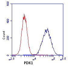
HL-60 cells were stained with 1 µg/mL PDK1 antibody (ab110335) (blue) or an equal amount of an isotype control antibody (red) and analyzed by flow cytometry.

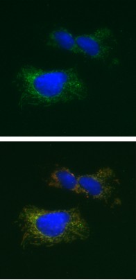
Immunocytochemistry image of PDK1 stained HeLa cells. The Upper image shows cells which were paraformaldehyde fixed (4%, 20 minutes) and Triton X-100 permeabilized (0.1%, 15 minutes). The cells were incubated with the PDK1 antibody (ab110335) at 2 µg/mL for 2 hours at room temperature or over night at 4°C. The secondary antibody was (green) Alexa Fluor® 488 goat anti-mouse IgG (H+L) at a 1/1000 dilution for 1 hour. 10% Goat serum was used as the blocking agent for all blocking steps. The target protein locates to the mitochondrial matrix. The lower image shows cells co-stained with an antibody against HSP60 (red), an enzyme also located in the mitochondrial matrix. The composite image shows an identical mitochondrial pattern for both antibodies indicated by merged orange color.
![All lanes : Anti-Mitochondrial Pyruvate dehydrogenase kinase 1 antibody [2H3AA11] (ab110335) at 1 µg/mlLane 1 : Human heart lysate at 10 µgLane 2 : Human liver lysate at 10 µgLane 3 : Recombinant Human PDK1 at 0.01 µgLane 4 : Recombinant Human PDK2 at 0.01 µgLane 5 : Recombinant Human PDK3 at 0.01 µgLane 6 : Recombinant Human PDK4 at 0.01 µg](http://www.bioprodhub.com/system/product_images/ab_products/2/sub_3/22846_Mitochondrial-Pyruvate-dehydrogenase-kinase-1-Primary-antibodies-ab110335-1.jpg)
![All lanes : Anti-Mitochondrial Pyruvate dehydrogenase kinase 1 antibody [2H3AA11] (ab110335) at 1 µg/mlLane 1 : HeLa cells incubated for 24 hours with vehicleLane 2 : HeLa cells incubated for 24 hours with 100 µM cobalt chloride (CoCl2) Lane 3 : HeLa cells incubated for 24 hours with 1 mM deferoxamine (DFO)Lysates/proteins at 30 µg per lane.](http://www.bioprodhub.com/system/product_images/ab_products/2/sub_3/22847_Mitochondrial-Pyruvate-dehydrogenase-kinase-1-Primary-antibodies-ab110335-8.jpg)

