Anti-Mps1 antibody [N1]
| Name | Anti-Mps1 antibody [N1] |
|---|---|
| Supplier | Abcam |
| Catalog | ab11108 |
| Prices | $401.00 |
| Sizes | 100 µl |
| Host | Mouse |
| Clonality | Monoclonal |
| Isotype | IgG1 |
| Clone | N1 |
| Applications | FC ICC/IF ICC/IF DB FA IP ELISA WB |
| Species Reactivities | Human |
| Antigen | Recombinant near full length protein (catalytically inactive - D663A), corresponding to amino acids 3-856 of Human Mps1 |
| Description | Mouse Monoclonal |
| Gene | TTK |
| Conjugate | Unconjugated |
| Supplier Page | Shop |
Product images
![All lanes : Anti-Mps1 antibody [N1] (ab11108) at 1 µg/mlLane 1 : HeLa (Human epithelial carcinoma cell line) Whole Cell Lysate Lane 2 : HeLa (Human epithelial carcinoma cell line) Nuclear Lysate Lane 3 : Jurkat (Human T cell lymphoblast-like cell line) Whole Cell Lysate Lane 4 : Jurkat (Human T cell lymphoblast-like cell line) Nuclear Lysate (ab14844)Lysates/proteins at 20 µg per lane.SecondaryGoat polyclonal to Mouse IgG - H&L - Pre-Adsorbed (HRP) at 1/3000 dilutiondeveloped using the ECL techniquePerformed under reducing conditions.](http://www.bioprodhub.com/system/product_images/ab_products/2/sub_3/24298_Mps1-Primary-antibodies-ab11108-12.jpg) All lanes : Anti-Mps1 antibody [N1] (ab11108) at 1 µg/mlLane 1 : HeLa (Human epithelial carcinoma cell line) Whole Cell Lysate Lane 2 : HeLa (Human epithelial carcinoma cell line) Nuclear Lysate Lane 3 : Jurkat (Human T cell lymphoblast-like cell line) Whole Cell Lysate Lane 4 : Jurkat (Human T cell lymphoblast-like cell line) Nuclear Lysate (ab14844)Lysates/proteins at 20 µg per lane.SecondaryGoat polyclonal to Mouse IgG - H&L - Pre-Adsorbed (HRP) at 1/3000 dilutiondeveloped using the ECL techniquePerformed under reducing conditions.
All lanes : Anti-Mps1 antibody [N1] (ab11108) at 1 µg/mlLane 1 : HeLa (Human epithelial carcinoma cell line) Whole Cell Lysate Lane 2 : HeLa (Human epithelial carcinoma cell line) Nuclear Lysate Lane 3 : Jurkat (Human T cell lymphoblast-like cell line) Whole Cell Lysate Lane 4 : Jurkat (Human T cell lymphoblast-like cell line) Nuclear Lysate (ab14844)Lysates/proteins at 20 µg per lane.SecondaryGoat polyclonal to Mouse IgG - H&L - Pre-Adsorbed (HRP) at 1/3000 dilutiondeveloped using the ECL techniquePerformed under reducing conditions.
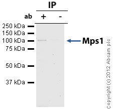 Mps1 was immunoprecipitated using 0.5mg Jurkat whole cell extract, 5µg of Mouse monoclonal to Mps1 and 50µl of protein G magnetic beads (+). No antibody was added to the control (-). The antibody was incubated under agitation with Protein G beads for 10min, Jurkat whole cell extract lysate diluted in RIPA buffer was added to each sample and incubated for a further 10min under agitation.Proteins were eluted by addition of 40µl SDS loading buffer and incubated for 10min at 70oC; 10µl of each sample was separated on a SDS PAGE gel, transferred to a nitrocellulose membrane, blocked with 5% BSA and probed with ab11108.Secondary: Goat polyclonal to mouse IgG light chain specific (HRP) at 1/5000 dilution.Band: 100kDa: Mps1
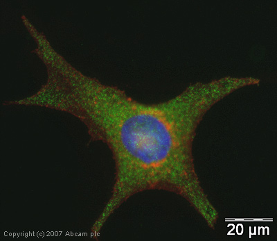 ICC/IF image of ab11108 stained human HeLa cells. The cells were PFA fixed (10 min), permabilised in TBS-T (20 min) and incubated with the antibody (ab11108, 1µg/ml) for 1h at room temperature. 1%BSA / 10% normal goat serum / 0.3M glycine was used to quench autofluorescence and block non-specific protein-protein interactions. The secondary antibody (green) was Alexa Fluor® 488 goat anti-mouse IgG (H+L) used at a 1/1000 dilution for 1h. Alexa Fluor® 594 WGA was used to label plasma membranes (red). DAPI was used to stain the cell nuclei (blue).
![Overlay histogram showing HeLa cells stained with ab11108 (red line). The cells were fixed with 80% methanol (5 min) and then permeabilized with 0.1% PBS-Tween for 20 min. The cells were then incubated in 1x PBS / 10% normal goat serum / 0.3M glycine to block non-specific protein-protein interactions followed by the antibody (ab11108, 2µg/1x106 cells) for 30 min at 22ºC. The secondary antibody used was DyLight® 488 goat anti-mouse IgG (H+L) (ab96879) at 1/500 dilution for 30 min at 22ºC. Isotype control antibody (black line) was mouse IgG1 [ICIGG1] (ab91353, 2µg/1x106 cells) used under the same conditions. Acquisition of >5,000 events was performed.](http://www.bioprodhub.com/system/product_images/ab_products/2/sub_3/24301_Mps1-Primary-antibodies-ab11108-13.jpg) Overlay histogram showing HeLa cells stained with ab11108 (red line). The cells were fixed with 80% methanol (5 min) and then permeabilized with 0.1% PBS-Tween for 20 min. The cells were then incubated in 1x PBS / 10% normal goat serum / 0.3M glycine to block non-specific protein-protein interactions followed by the antibody (ab11108, 2µg/1x106 cells) for 30 min at 22ºC. The secondary antibody used was DyLight® 488 goat anti-mouse IgG (H+L) (ab96879) at 1/500 dilution for 30 min at 22ºC. Isotype control antibody (black line) was mouse IgG1 [ICIGG1] (ab91353, 2µg/1x106 cells) used under the same conditions. Acquisition of >5,000 events was performed.
Overlay histogram showing HeLa cells stained with ab11108 (red line). The cells were fixed with 80% methanol (5 min) and then permeabilized with 0.1% PBS-Tween for 20 min. The cells were then incubated in 1x PBS / 10% normal goat serum / 0.3M glycine to block non-specific protein-protein interactions followed by the antibody (ab11108, 2µg/1x106 cells) for 30 min at 22ºC. The secondary antibody used was DyLight® 488 goat anti-mouse IgG (H+L) (ab96879) at 1/500 dilution for 30 min at 22ºC. Isotype control antibody (black line) was mouse IgG1 [ICIGG1] (ab91353, 2µg/1x106 cells) used under the same conditions. Acquisition of >5,000 events was performed.
![Anti-Mps1 antibody [N1] (ab11108) at 10 µg/ml + Active human Mps1 full length protein (ab89589) at 0.01 µgSecondaryGoat Anti-Mouse IgG H&L (HRP) preadsorbed (ab97040) at 1/5000 dilutiondeveloped using the ECL techniquePerformed under reducing conditions.Exposure time : 8 minutes](http://www.bioprodhub.com/system/product_images/ab_products/2/sub_3/24302_Mps1-Primary-antibodies-ab11108-16.jpg) Anti-Mps1 antibody [N1] (ab11108) at 10 µg/ml + Active human Mps1 full length protein (ab89589) at 0.01 µgSecondaryGoat Anti-Mouse IgG H&L (HRP) preadsorbed (ab97040) at 1/5000 dilutiondeveloped using the ECL techniquePerformed under reducing conditions.Exposure time : 8 minutes
Anti-Mps1 antibody [N1] (ab11108) at 10 µg/ml + Active human Mps1 full length protein (ab89589) at 0.01 µgSecondaryGoat Anti-Mouse IgG H&L (HRP) preadsorbed (ab97040) at 1/5000 dilutiondeveloped using the ECL techniquePerformed under reducing conditions.Exposure time : 8 minutes
Product References
Meta-analysis of the global gene expression profile of triple-negative breast - Meta-analysis of the global gene expression profile of triple-negative breast
Al-Ejeh F, Simpson PT, Sanus JM, Klein K, Kalimutho M, Shi W, Miranda M, Kutasovic J, Raghavendra A, Madore J, Reid L, Krause L, Chenevix-Trench G, Lakhani SR, Khanna KK. Oncogenesis. 2014 Apr 21;3:e100.
Chk2 prevents mitotic exit when the majority of kinetochores are unattached. - Chk2 prevents mitotic exit when the majority of kinetochores are unattached.
Petsalaki E, Zachos G. J Cell Biol. 2014 May 12;205(3):339-56.
Melanoma-associated mutations in protein phosphatase 6 cause chromosome - Melanoma-associated mutations in protein phosphatase 6 cause chromosome
Hammond D, Zeng K, Espert A, Bastos RN, Baron RD, Gruneberg U, Barr FA. J Cell Sci. 2013 Aug 1;126(Pt 15):3429-40.
Caspase-3-dependent mitotic checkpoint inactivation by the small-molecule - Caspase-3-dependent mitotic checkpoint inactivation by the small-molecule
Riffell JL, Janicke RU, Roberge M. Mol Cancer Ther. 2011 May;10(5):839-49.
B-Raf(V600E) signaling deregulates the mitotic spindle checkpoint through - B-Raf(V600E) signaling deregulates the mitotic spindle checkpoint through
Cui Y, Guadagno TM. Oncogene. 2008 May 15;27(22):3122-33. Epub 2007 Dec 10.
Kinetochore localization and microtubule interaction of the human spindle - Kinetochore localization and microtubule interaction of the human spindle
Stucke VM, Baumann C, Nigg EA. Chromosoma. 2004 Aug;113(1):1-15. Epub 2004 Jul 3.
Human Mps1 protein kinase is required for centrosome duplication and normal - Human Mps1 protein kinase is required for centrosome duplication and normal
Fisk HA, Mattison CP, Winey M. Proc Natl Acad Sci U S A. 2003 Dec 9;100(25):14875-80. Epub 2003 Dec 1.
Human Mps1 kinase is required for the spindle assembly checkpoint but not for - Human Mps1 kinase is required for the spindle assembly checkpoint but not for
Stucke VM, Sillje HH, Arnaud L, Nigg EA. EMBO J. 2002 Apr 2;21(7):1723-32.