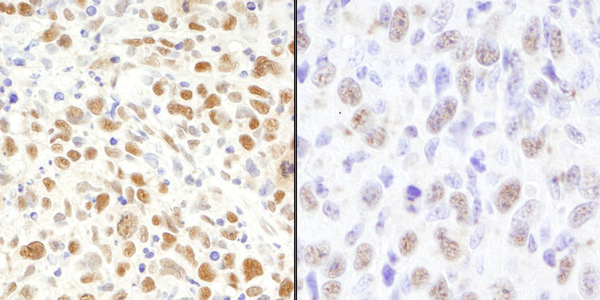
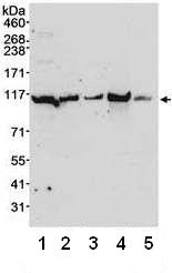
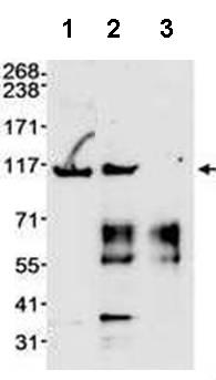
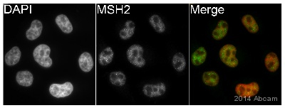
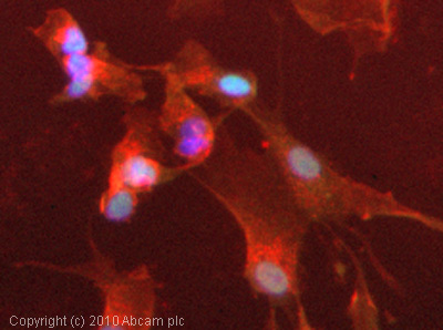
Anti-MSH2 antibody
| Name | Anti-MSH2 antibody |
|---|---|
| Supplier | Abcam |
| Catalog | ab70270 |
| Prices | $394.00 |
| Sizes | 100 µl |
| Host | Rabbit |
| Clonality | Polyclonal |
| Isotype | IgG |
| Applications | ICC/IF ICC/IF WB IP IHC-P |
| Species Reactivities | Mouse, Human, Guinea Pig, Bovine, Pig, Monkey, Ape, Monkey, Marmoset, Orangutan, Elephant |
| Antigen | Synthetic peptide: LEREQGEKII QEFLSKVKQM PFTEMSEENI TIKLKQLKAE VIAKNNSFVN EIISRIKVTT , corresponding to C terminal amino acids 875-934 of Human MSH2 Run BLAST with Run BLAST with |
| Description | Rabbit Polyclonal |
| Gene | MSH2 |
| Conjugate | Unconjugated |
| Supplier Page | Shop |
Product images
Product References
X inactivation plays a major role in the gender bias in somatic expansion in a - X inactivation plays a major role in the gender bias in somatic expansion in a
Adihe Lokanga R, Zhao XN, Entezam A, Usdin K. Hum Mol Genet. 2014 Sep 15;23(18):4985-94.
Msh2 acts in medium-spiny striatal neurons as an enhancer of CAG instability and - Msh2 acts in medium-spiny striatal neurons as an enhancer of CAG instability and
Kovalenko M, Dragileva E, St Claire J, Gillis T, Guide JR, New J, Dong H, Kucherlapati R, Kucherlapati MH, Ehrlich ME, Lee JM, Wheeler VC. PLoS One. 2012;7(9):e44273.
