Anti-MSH6 antibody [EPR3945]
| Name | Anti-MSH6 antibody [EPR3945] |
|---|---|
| Supplier | Abcam |
| Catalog | ab92471 |
| Prices | $401.00 |
| Sizes | 100 µl |
| Host | Rabbit |
| Clonality | Monoclonal |
| Isotype | IgG |
| Clone | EPR3945 |
| Applications | WB IHC-P ICC/IF ICC/IF |
| Species Reactivities | Mouse, Rat, Human |
| Antigen | Synthetic peptide (the amino acid sequence is considered to be commercially sensitive) corresponding to Human MSH6 aa 1-100 (N terminal) |
| Description | Rabbit Monoclonal |
| Gene | MSH6 |
| Conjugate | Unconjugated |
| Supplier Page | Shop |
Product images
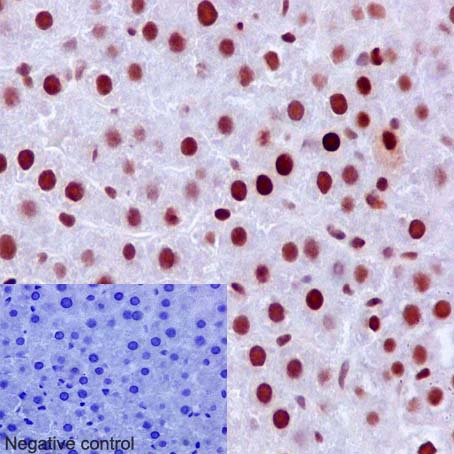 Immunohistochemical staining of paraffin embedded rat liver with purified ab92471 at a dilution of 1/500. A pre-diluted HRP polymer for rabbit/mouse IgG was used as the secondary antibody and the sample was counter-stained with hematoxylin. Antigen retrieval was perfomed using Tris-EDTA buffer, pH 9.0. PBS was used instead of the primary antibody as the negative control, and is shown in the inset.
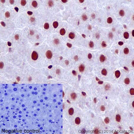 Immunohistochemical staining of paraffin embedded rat liver with unpurified ab92471 at a dilution of 1/150. A pre-diluted HRP polymer for rabbit/mouse IgG was used as the secondary antibody and the sample was counter-stained with hematoxylin. Antigen retrieval was perfomed using Tris-EDTA buffer, pH 9.0. PBS was used instead of the primary antibody as the negative control, and is shown in the inset.
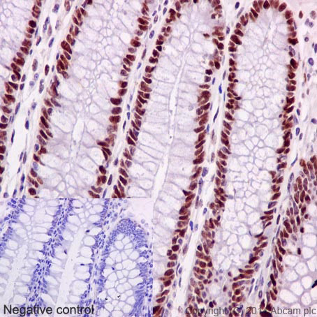 Immunohistochemical staining of paraffin embedded human colon with unpurified ab92471 at a dilution of 1/500. A pre-diluted HRP polymer for rabbit/mouse IgG was used as the secondary antibody and the sample was counter-stained with hematoxylin. Antigen retrieval was perfomed using Tris-EDTA buffer, pH 9.0. PBS was used instead of the primary antibody as the negative control, and is shown in the inset.
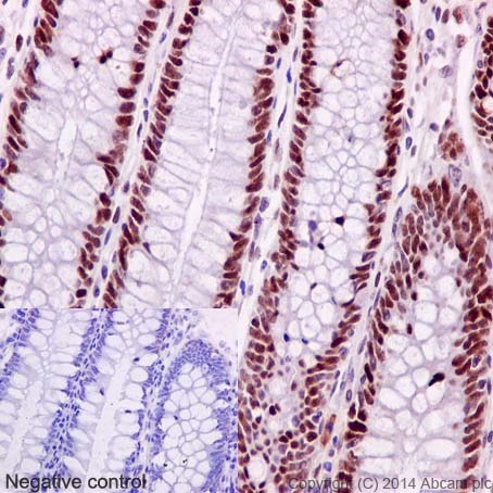 Immunohistochemical staining of paraffin embedded human colon with unpurified ab92471 at a dilution of 1/150. A pre-diluted HRP polymer for rabbit/mouse IgG was used as the secondary antibody and the sample was counter-stained with hematoxylin. Antigen retrieval was perfomed using Tris-EDTA buffer, pH 9.0. PBS was used instead of the primary antibody as the negative control, and is shown in the inset.
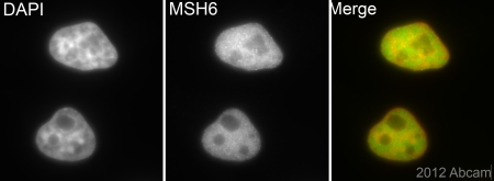
 Immunofluorescent staining of HeLa cells (fixed in 4% PFA, permeabilized with 0.1% Triton X 100) using purified ab92471 at a dilution of 1/250. An Alexa Fluor® 488 goat anti-rabbit antibody was used as the secondary at a dilution of 1/500 and the cells were counter stained with DAPI. The negative control is shown in the bottom right hand panel - for the negative control, Alex Fluor® 488 goat anti-mouse was used at a dilution of 1/500.
![Anti-MSH6 antibody [EPR3945] (ab92471) at 1/1000 dilution (purified) + Rat brain at 10 µgSecondaryHRP goat anti-rabbit (H+L) at 1/1000 dilution](http://www.bioprodhub.com/system/product_images/ab_products/2/sub_3/25081_ab92471-238045-92471-WB-3.jpg) Anti-MSH6 antibody [EPR3945] (ab92471) at 1/1000 dilution (purified) + Rat brain at 10 µgSecondaryHRP goat anti-rabbit (H+L) at 1/1000 dilution
Anti-MSH6 antibody [EPR3945] (ab92471) at 1/1000 dilution (purified) + Rat brain at 10 µgSecondaryHRP goat anti-rabbit (H+L) at 1/1000 dilution
 purified at 1/6000 dilution + SW480 cell lysate at 10 µgSecondaryHRP goat anti-rabbit (H+L) at 1/1000 dilution
![Anti-MSH6 antibody [EPR3945] (ab92471) at 1/2000 dilution (unpurified) + SW480 cell lysate at 10 µgSecondaryHRP goat anti-rabbit (H+L) at 1/1000 dilution](http://www.bioprodhub.com/system/product_images/ab_products/2/sub_3/25083_ab92471-238043-92471-WB-1.jpg) Anti-MSH6 antibody [EPR3945] (ab92471) at 1/2000 dilution (unpurified) + SW480 cell lysate at 10 µgSecondaryHRP goat anti-rabbit (H+L) at 1/1000 dilution
Anti-MSH6 antibody [EPR3945] (ab92471) at 1/2000 dilution (unpurified) + SW480 cell lysate at 10 µgSecondaryHRP goat anti-rabbit (H+L) at 1/1000 dilution
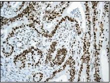 Unpurified ab92471, at a 1/100 dilution, detecting MSH6 in paraffin embedded Human colonic adenocarcinoma tissue by immunohistochemistry. Detection used HRP conjugated anti rabbit antibody.
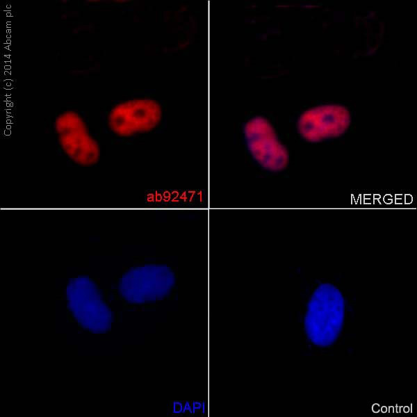
 Unpurified ab92471 (1/500) staining MSH6 in asynchronous HeLa cells (green). Cells were fixed in paraformaldehyde, permeabilised with 0.5% Triton X100/PBS and counterstained with DAPI in order to highlight the nucleus (red). For further experimental details please see abreview.See Abreview
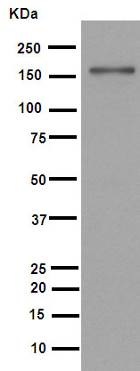
![All lanes : Anti-MSH6 antibody [EPR3945] (ab92471) at 1/1000 dilution (unpurified)Lane 1 : A431 cell lysateLane 2 : HeLa cell lysateLane 3 : SW480 cell lysateLysates/proteins at 10 µg per lane.SecondaryHRP labelled goat anti-rabbit antibody at 1/2000 dilution](http://www.bioprodhub.com/system/product_images/ab_products/2/sub_3/25086_MSH6-Primary-antibodies-ab92471-1.jpg) All lanes : Anti-MSH6 antibody [EPR3945] (ab92471) at 1/1000 dilution (unpurified)Lane 1 : A431 cell lysateLane 2 : HeLa cell lysateLane 3 : SW480 cell lysateLysates/proteins at 10 µg per lane.SecondaryHRP labelled goat anti-rabbit antibody at 1/2000 dilution
All lanes : Anti-MSH6 antibody [EPR3945] (ab92471) at 1/1000 dilution (unpurified)Lane 1 : A431 cell lysateLane 2 : HeLa cell lysateLane 3 : SW480 cell lysateLysates/proteins at 10 µg per lane.SecondaryHRP labelled goat anti-rabbit antibody at 1/2000 dilution
 Equilibrium disassociation constant (KD)Learn more about KD Click here to learn more about KD
Product References
Immune chaperone gp96 drives the contributions of macrophages to inflammatory - Immune chaperone gp96 drives the contributions of macrophages to inflammatory
Morales C, Rachidi S, Hong F, Sun S, Ouyang X, Wallace C, Zhang Y, Garret-Mayer E, Wu J, Liu B, Li Z. Cancer Res. 2014 Jan 15;74(2):446-59.
Traditional serrated adenoma has two pathways of neoplastic progression that are - Traditional serrated adenoma has two pathways of neoplastic progression that are
Tsai JH, Liau JY, Lin YL, Lin LI, Cheng YC, Cheng ML, Jeng YM. Mod Pathol. 2014 Oct;27(10):1375-85.
Heterogenous mismatch-repair status in colorectal cancer. - Heterogenous mismatch-repair status in colorectal cancer.
Joost P, Veurink N, Holck S, Klarskov L, Bojesen A, Harbo M, Baldetorp B, Rambech E, Nilbert M. Diagn Pathol. 2014 Jun 26;9:126.
Aberrant expression of annexin A10 is closely related to gastric phenotype in - Aberrant expression of annexin A10 is closely related to gastric phenotype in
Tsai JH, Lin YL, Cheng YC, Chen CC, Lin LI, Tseng LH, Cheng ML, Liau JY, Jeng YM. Mod Pathol. 2015 Feb;28(2):268-78.
MMR deficiency is common in high-grade endometrioid carcinomas and is associated - MMR deficiency is common in high-grade endometrioid carcinomas and is associated
Nelson GS, Pink A, Lee S, Han G, Morris D, Ogilvie T, Duggan MA, Kobel M. Gynecol Oncol. 2013 Nov;131(2):309-14.
Efficient and reproducible identification of mismatch repair deficient colon - Efficient and reproducible identification of mismatch repair deficient colon
Joost P, Bendahl PO, Halvarsson B, Rambech E, Nilbert M. BMC Clin Pathol. 2013 Dec 17;13(1):33.
A cohort study of the prognostic and treatment predictive value of SATB2 - A cohort study of the prognostic and treatment predictive value of SATB2
Eberhard J, Gaber A, Wangefjord S, Nodin B, Uhlen M, Ericson Lindquist K, Jirstrom K. Br J Cancer. 2012 Feb 28;106(5):931-8.
Acquired resistance to temozolomide in glioma cell lines: molecular mechanisms - Acquired resistance to temozolomide in glioma cell lines: molecular mechanisms
Zhang J, Stevens MF, Laughton CA, Madhusudan S, Bradshaw TD. Oncology. 2010;78(2):103-14.