![Anti-Myelin Basic Protein antibody [7D2] (ab78157) at 1/10000 dilution + Crude rat spinal cord homogenate](http://www.bioprodhub.com/system/product_images/ab_products/2/sub_3/26440_ab78157-1.jpg)
Anti-Myelin Basic Protein antibody [7D2] (ab78157) at 1/10000 dilution + Crude rat spinal cord homogenate
![Lane 1 : Anti-Myelin Basic Protein antibody [7G7] (ab78156) at 1/10000 dilutionLane 2 : Anti-Myelin Basic Protein antibody [7D2] (ab78157) at 1/10000 dilutionLane 1 : Crude rat spinal cord homogenateLane 2 : Crude rat spinal cord homogenate](http://www.bioprodhub.com/system/product_images/ab_products/2/sub_3/26441_ab78157-3.jpg)
Lane 1 : Anti-Myelin Basic Protein antibody [7G7] (ab78156) at 1/10000 dilutionLane 2 : Anti-Myelin Basic Protein antibody [7D2] (ab78157) at 1/10000 dilutionLane 1 : Crude rat spinal cord homogenateLane 2 : Crude rat spinal cord homogenate

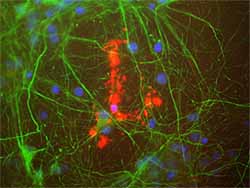
ab78157 (red) staining Myelin Basic Protein in rat mixed neuron/glial cultures. Also stained with a chicken antibody to neurofilament NF-L (green). Blue is a DNA stain. Note that ab78157 stains an oligodendrocyte and some membrane shed from this cell. Other cells in the field include neurons, astrocytes, microglia and fibroblasts, all of which are completely negative for Myelin Basic Protein, though the neuronal processes can be seen with the NF-L antibody.
![Overlay histogram showing SH-SY5Y cells stained with ab78157 (red line). The cells were fixed with 80% methanol (5 min) and then permeabilized with 0.1% PBS-Tween for 20 min. The cells were then incubated in 1x PBS / 10% normal goat serum / 0.3M glycine to block non-specific protein-protein interactions followed by the antibody (ab78157, 1µg/1x106 cells) for 30 min at 22ºC. The secondary antibody used was DyLight® 488 goat anti-mouse IgG (H+L) (ab96879) at 1/500 dilution for 30 min at 22ºC. Isotype control antibody (black line) was mouse IgG1 [ICIGG1] (ab91353, 2µg/1x106 cells) used under the same conditions. Acquisition of >5,000 events was performed. This antibody gave a positive signal in SH-SY5Y cells fixed with 4% paraformaldehyde (10 min)/permeabilized with 0.1% PBS-Tween for 20 min used under the same conditions.](http://www.bioprodhub.com/system/product_images/ab_products/2/sub_3/26443_Myelin-Basic-Protein-Primary-antibodies-ab78157-1.jpg)
Overlay histogram showing SH-SY5Y cells stained with ab78157 (red line). The cells were fixed with 80% methanol (5 min) and then permeabilized with 0.1% PBS-Tween for 20 min. The cells were then incubated in 1x PBS / 10% normal goat serum / 0.3M glycine to block non-specific protein-protein interactions followed by the antibody (ab78157, 1µg/1x106 cells) for 30 min at 22ºC. The secondary antibody used was DyLight® 488 goat anti-mouse IgG (H+L) (ab96879) at 1/500 dilution for 30 min at 22ºC. Isotype control antibody (black line) was mouse IgG1 [ICIGG1] (ab91353, 2µg/1x106 cells) used under the same conditions. Acquisition of >5,000 events was performed. This antibody gave a positive signal in SH-SY5Y cells fixed with 4% paraformaldehyde (10 min)/permeabilized with 0.1% PBS-Tween for 20 min used under the same conditions.
![Anti-Myelin Basic Protein antibody [7D2] (ab78157) at 1/10000 dilution + Crude rat spinal cord homogenate](http://www.bioprodhub.com/system/product_images/ab_products/2/sub_3/26440_ab78157-1.jpg)
![Lane 1 : Anti-Myelin Basic Protein antibody [7G7] (ab78156) at 1/10000 dilutionLane 2 : Anti-Myelin Basic Protein antibody [7D2] (ab78157) at 1/10000 dilutionLane 1 : Crude rat spinal cord homogenateLane 2 : Crude rat spinal cord homogenate](http://www.bioprodhub.com/system/product_images/ab_products/2/sub_3/26441_ab78157-3.jpg)

![Overlay histogram showing SH-SY5Y cells stained with ab78157 (red line). The cells were fixed with 80% methanol (5 min) and then permeabilized with 0.1% PBS-Tween for 20 min. The cells were then incubated in 1x PBS / 10% normal goat serum / 0.3M glycine to block non-specific protein-protein interactions followed by the antibody (ab78157, 1µg/1x106 cells) for 30 min at 22ºC. The secondary antibody used was DyLight® 488 goat anti-mouse IgG (H+L) (ab96879) at 1/500 dilution for 30 min at 22ºC. Isotype control antibody (black line) was mouse IgG1 [ICIGG1] (ab91353, 2µg/1x106 cells) used under the same conditions. Acquisition of >5,000 events was performed. This antibody gave a positive signal in SH-SY5Y cells fixed with 4% paraformaldehyde (10 min)/permeabilized with 0.1% PBS-Tween for 20 min used under the same conditions.](http://www.bioprodhub.com/system/product_images/ab_products/2/sub_3/26443_Myelin-Basic-Protein-Primary-antibodies-ab78157-1.jpg)