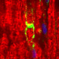
A tissue section through an adult sciatic nerve. Myelin Protein Zero (green staining) can be seen in the myelin and Schwann cell processes surrounding the nodes of Ranvier. In this photomicrograph, rabbit antibodies against LAMP (lysozome-associated membrane glycoprotein) (red staining) serves as the counterstain, and DAPI (blue staining) allows visualization of nuclei.

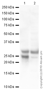
All lanes : Anti-Myelin Protein Zero antibody (ab39375) at 1 µg/mlLane 1 : Spinal Cord (Human) Tissue Lysate - adult normal tissue (ab29188)Lane 2 : Spinal Cord (Mouse) Tissue Lysate Lysates/proteins at 10 µg per lane.SecondaryGoat polyclonal Secondary Antibody to Chicken IgY - H&L (HRP) at 1/3000 dilutiondeveloped using the ECL techniquePerformed under reducing conditions.

