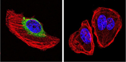
Immunofluorescent analysis of formaldehyde-fixed U251 cells labeling Neurofibromin with ab178323 at 1/20 dilution (green, left image) or a control (right image) followed with a DyLight-488 conjugated secondary antibody. F-Actin staining with Phalloidin (red) and nuclei with DAPI (blue).
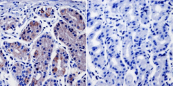
Immunohistochemical analysis of deparaffinized Human stomach tissue labeling Neurofibromin with ab178323 at 1/20 dilution or without primary antibody (negative control). Detection was performed using a biotin-conjugated secondary antibody and SA-HRP, followed by colorimetric detection using DAB. Tissues were counterstained with hematoxylin.
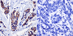
Immunohistochemical analysis of deparaffinized Human breast carcinoma tissue labeling Neurofibromin with ab178323 at 1/20 dilution or without primary antibody (negative control). Detection was performed using a biotin-conjugated secondary antibody and SA-HRP, followed by colorimetric detection using DAB. Tissues were counterstained with hematoxylin.
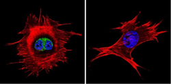
Immunofluorescent analysis of formaldehyde-fixed NIH 3T3 cells labeling Neurofibromin with ab178323 at 1/20 dilution (green, left image) or a control (right image) followed with a DyLight-488 conjugated secondary antibody. F-Actin staining with Phalloidin (red) and nuclei with DAPI (blue).
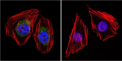
Immunofluorescent analysis of formaldehyde-fixed HeLa cells labeling Neurofibromin with ab178323 at 1/20 dilution (green, left image) or a control (right image) followed with a DyLight-488 conjugated secondary antibody. F-Actin staining with Phalloidin (red) and nuclei with DAPI (blue).


