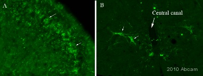
IHC-FoFr image of Neuroligin 2 (ab36602) staining on Rat Spinal Cord. The sections used came from animals perfused fixed with Paraformaldehyde 4%, in phosphate buffer 0.2M. Following postfixation in the same fixative overnight, the tissues were cryoprotected in sucrose 30% overnight. Tissues were then cut using a cryostat and the immunostainings were preformed using the ‘free floating’ technique. Image A shows the staining observed at the level of the lateral part of the dorsal horn, showing stained lamina II neurons (arrows). Image B shows the staining observed lamina X. See Abreview
