![All lanes : Anti-NGF antibody [EP1320Y] (ab52918) at 1/1000 dilution (purified)Lane 1 : Human fetal brain lysateLane 2 : Human fetal thymus lysateLane 3 : HeLa cell lysateLysates/proteins at 20 µg per lane.SecondaryHRP conjugated goat anti-rabbit IgG (H+L) at 1/1000 dilution](http://www.bioprodhub.com/system/product_images/ab_products/2/sub_4/176_ab52918-4-ab52918WB1.jpg)
All lanes : Anti-NGF antibody [EP1320Y] (ab52918) at 1/1000 dilution (purified)Lane 1 : Human fetal brain lysateLane 2 : Human fetal thymus lysateLane 3 : HeLa cell lysateLysates/proteins at 20 µg per lane.SecondaryHRP conjugated goat anti-rabbit IgG (H+L) at 1/1000 dilution
![Anti-NGF antibody [EP1320Y] (ab52918) at 1/1000 dilution (purified) + Mouse thyroid lysate at 20 µgSecondaryHRP conjugated goat anti-rabbit IgG (H+L) at 1/1000 dilution](http://www.bioprodhub.com/system/product_images/ab_products/2/sub_4/177_ab52918-6-ab52918WB3.jpg)
Anti-NGF antibody [EP1320Y] (ab52918) at 1/1000 dilution (purified) + Mouse thyroid lysate at 20 µgSecondaryHRP conjugated goat anti-rabbit IgG (H+L) at 1/1000 dilution
![Anti-NGF antibody [EP1320Y] (ab52918) at 1/2000 dilution (purified) + Rat thyroid lysate at 20 µgSecondaryHRP conjugated goat anti-rabbit IgG (H+L) at 1/1000 dilution](http://www.bioprodhub.com/system/product_images/ab_products/2/sub_4/178_ab52918-5-ab52918WB2.jpg)
Anti-NGF antibody [EP1320Y] (ab52918) at 1/2000 dilution (purified) + Rat thyroid lysate at 20 µgSecondaryHRP conjugated goat anti-rabbit IgG (H+L) at 1/1000 dilution

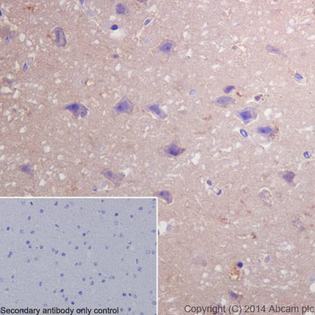
Immunohistochemical staining of paraffin embedded human cerebral cortex with purified ab52918 at a working dilution of 1 in 250. The secondary antibody used is ab97051 Goat Anti-Rabbit IgG H&L (HRP) at a dilution of 1/500. The sample is counter-stained with hematoxylin. Antigen retrieval was perfomed using Tris-EDTA buffer, pH 9.0. PBS was used instead of the primary antibody as the negative control, and is shown in the inset.

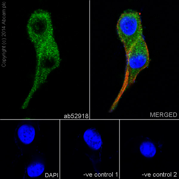
Immunofluorescence staining of U87-MG cells with purified ab52918 at a working dilution of 1 in 300, counter-stained with DAPI. Tubulin was stained with mouse anti-tubulin at a dilution of 1/1000 (ab7291) and Alexa Fluor® 594 goat anti-mouse at a dilution of 1/500 (ab150120) . The secondary antibody was ab150077 Alexa Fluor® 488 goat anti rabbit, used at a dilution of 1 in 500. The cells were fixed in 4% PFA and permeabilized using 0.1% Triton X 100. The negative controls are shown in the bottom middle and right hand panels - for the first negative control, purified ab52918 was used at a dilution of 1/200 followed by an Alexa Fluor® 555 goat anti-mouse antibody at a dilution of 1/500 and for the second negative control mouse primary antibody (ab7291) and anti-rabbit secondary antibody (ab15007) were used.
![Anti-NGF antibody [EP1320Y] (ab52918) at 1/500 dilution (unpurified) + fetal thyroid tissue at 10 µgSecondaryGoat anti rabbit IgG HRP antibody at 1/2000 dilution](http://www.bioprodhub.com/system/product_images/ab_products/2/sub_4/181_ab52918wb.jpg)
Anti-NGF antibody [EP1320Y] (ab52918) at 1/500 dilution (unpurified) + fetal thyroid tissue at 10 µgSecondaryGoat anti rabbit IgG HRP antibody at 1/2000 dilution

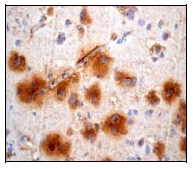
Immunohistochemical staining of paraffin embedded human brain using unpurified b52918 at 1/50-1/100 dilution.Ab52918 at 1/50-1/100 dilution staining human brain; paraffin embedded.

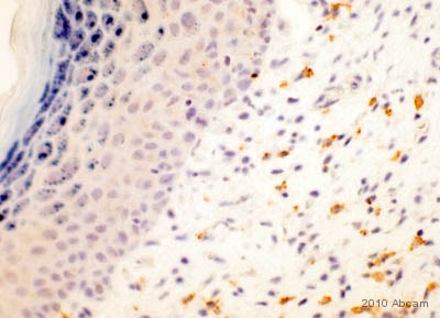
Immunohistochemical staining of NGF mouse tissue sections (formalin/ PFA-fixed paraffin-embedded tissue sections) with unpurified ab52918. The sections were formaldehyde fixed, subjected to heat mediated antigen retrieval and blocked for 10 minutes at 25°C. The primary antibody was diluted 1/50 and incubated with the sample for 1 hour at 25°C. An HRP polymer anti-rabbit IgG system was used undiluted, as the secondary antibody. See Abreview
![All lanes : Anti-NGF antibody [EP1320Y] (ab52918) at 1/1000 dilution (purified)Lane 1 : Human fetal brain lysateLane 2 : Human fetal thymus lysateLane 3 : HeLa cell lysateLysates/proteins at 20 µg per lane.SecondaryHRP conjugated goat anti-rabbit IgG (H+L) at 1/1000 dilution](http://www.bioprodhub.com/system/product_images/ab_products/2/sub_4/176_ab52918-4-ab52918WB1.jpg)
![Anti-NGF antibody [EP1320Y] (ab52918) at 1/1000 dilution (purified) + Mouse thyroid lysate at 20 µgSecondaryHRP conjugated goat anti-rabbit IgG (H+L) at 1/1000 dilution](http://www.bioprodhub.com/system/product_images/ab_products/2/sub_4/177_ab52918-6-ab52918WB3.jpg)
![Anti-NGF antibody [EP1320Y] (ab52918) at 1/2000 dilution (purified) + Rat thyroid lysate at 20 µgSecondaryHRP conjugated goat anti-rabbit IgG (H+L) at 1/1000 dilution](http://www.bioprodhub.com/system/product_images/ab_products/2/sub_4/178_ab52918-5-ab52918WB2.jpg)


![Anti-NGF antibody [EP1320Y] (ab52918) at 1/500 dilution (unpurified) + fetal thyroid tissue at 10 µgSecondaryGoat anti rabbit IgG HRP antibody at 1/2000 dilution](http://www.bioprodhub.com/system/product_images/ab_products/2/sub_4/181_ab52918wb.jpg)

