![All lanes : Anti-NIT1 antibody [4D12] (ab118158) at 1/1000 dilutionLane 1 : HEK293T cell lysate transfected with pCMV6-ENTRY controlLane 2 : HEK293T cell lysate transfected with pCMV6-ENTRY NIT1 cDNALysates/proteins at 5 µg per lane.](http://www.bioprodhub.com/system/product_images/ab_products/2/sub_4/484_NIT1-Primary-antibodies-ab118158-2.jpg)
All lanes : Anti-NIT1 antibody [4D12] (ab118158) at 1/1000 dilutionLane 1 : HEK293T cell lysate transfected with pCMV6-ENTRY controlLane 2 : HEK293T cell lysate transfected with pCMV6-ENTRY NIT1 cDNALysates/proteins at 5 µg per lane.
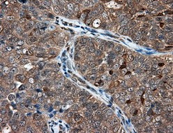
Immunohistochemical staining of paraffin-embedded Adenocarcinoma of ovary tissue using ab118158 at a dilution of 1/50.
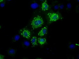
Immunofluorescent staining of COS7 cells transiently transfected with NIT1 using ab118158 at a dilution of 1/100.
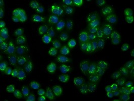
Immunofluorescent staining of HT29 cells using ab118158 at a dilution of 1/100.
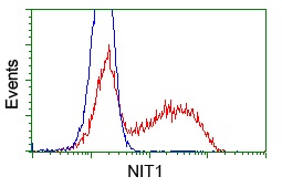
HEK293T cells transfected with either pCMV6-ENTRY NIT1 (Red) or empty vector control plasmid (Blue) were immunostained with ab118158 at a dilution of 1/100, and then analyzed by flow cytometry.