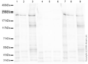
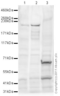
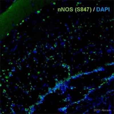
Anti-nNOS (neuronal) (phospho S847) antibody
| Name | Anti-nNOS (neuronal) (phospho S847) antibody |
|---|---|
| Supplier | Abcam |
| Catalog | ab16650 |
| Prices | $392.00 |
| Sizes | 100 µg |
| Host | Rabbit |
| Clonality | Polyclonal |
| Isotype | IgG |
| Applications | IHC-F IHC-F WB |
| Species Reactivities | Mouse, Rat, Fish, Rabbit, Human, Xenopus, Zebrafish |
| Antigen | Synthetic peptide conjugated to KLH derived from within residues 800 - 900 of Mouse nNOS (neuronal), phosphorylated at S847 |
| Blocking Peptide | Mouse nNOS (neuronal) (phospho S847) peptide |
| Description | Rabbit Polyclonal |
| Gene | NOS1 |
| Conjugate | Unconjugated |
| Supplier Page | Shop |
Product images
Product References
beta3-adrenoreceptor stimulation protects against myocardial infarction injury - beta3-adrenoreceptor stimulation protects against myocardial infarction injury
Niu X, Zhao L, Li X, Xue Y, Wang B, Lv Z, Chen J, Sun D, Zheng Q. PLoS One. 2014 Jun 9;9(6):e98713.
RGSZ2 binds to the neural nitric oxide synthase PDZ domain to regulate mu-opioid - RGSZ2 binds to the neural nitric oxide synthase PDZ domain to regulate mu-opioid
Garzon J, Rodriguez-Munoz M, Vicente-Sanchez A, Bailon C, Martinez-Murillo R, Sanchez-Blazquez P. Antioxid Redox Signal. 2011 Aug 15;15(4):873-87.
Role of neuronal nitric-oxide synthase in estrogen-induced relaxation in rat - Role of neuronal nitric-oxide synthase in estrogen-induced relaxation in rat
Lekontseva O, Chakrabarti S, Jiang Y, Cheung CC, Davidge ST. J Pharmacol Exp Ther. 2011 Nov;339(2):367-75.
Sigma receptor ligand 4-phenyl-1-(4-phenylbutyl)-piperidine modulates neuronal - Sigma receptor ligand 4-phenyl-1-(4-phenylbutyl)-piperidine modulates neuronal
Yang ZJ, Carter EL, Torbey MT, Martin LJ, Koehler RC. Exp Neurol. 2010 Jan;221(1):166-74.
Prolactin promotes oxytocin and vasopressin release by activating neuronal nitric - Prolactin promotes oxytocin and vasopressin release by activating neuronal nitric
Vega C, Moreno-Carranza B, Zamorano M, Quintanar-Stephano A, Mendez I, Thebault S, Martinez de la Escalera G, Clapp C. Am J Physiol Regul Integr Comp Physiol. 2010 Dec;299(6):R1701-8. doi:
Mu-opioid receptors transiently activate the Akt-nNOS pathway to produce - Mu-opioid receptors transiently activate the Akt-nNOS pathway to produce
Sanchez-Blazquez P, Rodriguez-Munoz M, Garzon J. PLoS One. 2010 Jun 23;5(6):e11278.
Coupling between neuronal nitric oxide synthase and glutamate receptor 6-mediated - Coupling between neuronal nitric oxide synthase and glutamate receptor 6-mediated
Yu HM, Xu J, Li C, Zhou C, Zhang F, Han D, Zhang GY. Neuroscience. 2008 Sep 9;155(4):1120-32.


