Anti-NUMB antibody
| Name | Anti-NUMB antibody |
|---|---|
| Supplier | Abcam |
| Catalog | ab14140 |
| Prices | $400.00 |
| Sizes | 100 µg |
| Host | Rabbit |
| Clonality | Polyclonal |
| Isotype | IgG |
| Applications | IHC-P ICC/IF ICC/IF IHC-F IHC WB IP |
| Species Reactivities | Mouse, Rat, Human, Zebrafish, Chicken, Monkey, Primate |
| Antigen | Synthetic peptide derived from residues 600 to the C-terminus of Human NUMB |
| Blocking Peptide | Human NUMB peptide |
| Description | Rabbit Polyclonal |
| Gene | NUMB |
| Conjugate | Unconjugated |
| Supplier Page | Shop |
Product images
Product References
Numb family proteins are essential for cardiac morphogenesis and progenitor - Numb family proteins are essential for cardiac morphogenesis and progenitor
Zhao C, Guo H, Li J, Myint T, Pittman W, Yang L, Zhong W, Schwartz RJ, Schwarz JJ, Singer HA, Tallquist MD, Wu M. Development. 2014 Jan;141(2):281-95.
Satb1 regulates the self-renewal of hematopoietic stem cells by promoting - Satb1 regulates the self-renewal of hematopoietic stem cells by promoting
Will B, Vogler TO, Bartholdy B, Garrett-Bakelman F, Mayer J, Barreyro L, Pandolfi A, Todorova TI, Okoye-Okafor UC, Stanley RF, Bhagat TD, Verma A, Figueroa ME, Melnick A, Roth M, Steidl U. Nat Immunol. 2013 May;14(5):437-45.
Expression and function of NUMB in odontogenesis. - Expression and function of NUMB in odontogenesis.
Li H, Ramachandran A, Gao Q, Ravindran S, Song Y, Evans C, George A. Biomed Res Int. 2013;2013:182965.
Angiotensin II inhibits satellite cell proliferation and prevents skeletal muscle - Angiotensin II inhibits satellite cell proliferation and prevents skeletal muscle
Yoshida T, Galvez S, Tiwari S, Rezk BM, Semprun-Prieto L, Higashi Y, Sukhanov S, Yablonka-Reuveni Z, Delafontaine P. J Biol Chem. 2013 Aug 16;288(33):23823-32.
Interleukin-10 regulates progenitor differentiation and modulates neurogenesis in - Interleukin-10 regulates progenitor differentiation and modulates neurogenesis in
Perez-Asensio FJ, Perpina U, Planas AM, Pozas E. J Cell Sci. 2013 Sep 15;126(Pt 18):4208-19.
Macrophage-derived Wnt opposes Notch signaling to specify hepatic progenitor cell - Macrophage-derived Wnt opposes Notch signaling to specify hepatic progenitor cell
Boulter L, Govaere O, Bird TG, Radulescu S, Ramachandran P, Pellicoro A, Ridgway RA, Seo SS, Spee B, Van Rooijen N, Sansom OJ, Iredale JP, Lowell S, Roskams T, Forbes SJ. Nat Med. 2012 Mar 4;18(4):572-9.
Numb expression and asymmetric versus symmetric cell division in distal embryonic - Numb expression and asymmetric versus symmetric cell division in distal embryonic
El-Hashash AH, Warburton D. J Histochem Cytochem. 2012 Sep;60(9):675-82.
The adaptor-associated kinase 1, AAK1, is a positive regulator of the Notch - The adaptor-associated kinase 1, AAK1, is a positive regulator of the Notch
Gupta-Rossi N, Ortica S, Meas-Yedid V, Heuss S, Moretti J, Olivo-Marin JC, Israel A. J Biol Chem. 2011 May 27;286(21):18720-30.
Functional dicer is necessary for appropriate specification of radial glia during - Functional dicer is necessary for appropriate specification of radial glia during
Nowakowski TJ, Mysiak KS, Pratt T, Price DJ. PLoS One. 2011;6(8):e23013.
Epicardial spindle orientation controls cell entry into the myocardium. - Epicardial spindle orientation controls cell entry into the myocardium.
Wu M, Smith CL, Hall JA, Lee I, Luby-Phelps K, Tallquist MD. Dev Cell. 2010 Jul 20;19(1):114-25.


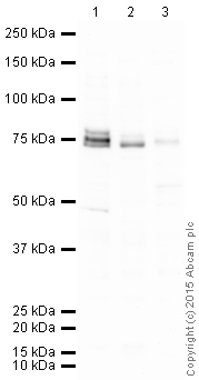
![NUMB was immunoprecipitated using 0.5mg Mouse Brain whole tissue lysate, 5µg of Rabbit polyclonal to NUMB and 50µl of protein G magnetic beads (+). No antibody was added to the control (-). The antibody was incubated under agitation with Protein G beads for 10min, Mouse Brain whole tissue lysate lysate diluted in RIPA buffer was added to each sample and incubated for a further 10min under agitation.Proteins were eluted by addition of 40µl SDS loading buffer and incubated for 10min at 70oC; 10µl of each sample was separated on a SDS PAGE gel, transferred to a nitrocellulose membrane, blocked with 5% BSA and probed with ab14140.Secondary: Mouse monoclonal [SB62a] Secondary Antibody to Rabbit IgG light chain (HRP) (ab99697).Band: 75kDa: NUMB.](http://www.bioprodhub.com/system/product_images/ab_products/2/sub_4/2925_NUMB-Primary-antibodies-ab14140-21.jpg)


