![All lanes : Anti-Olig2 antibody [EPR2673] (ab109186) at 1/2000 dilution (purified)Lane 1 : Mouse brain lysateLane 2 : Mouse brain lysateLysates/proteins at 20 µg per lane.SecondaryHRP goat anti-rabbit (H+L) at 1/1000 dilution](http://www.bioprodhub.com/system/product_images/ab_products/2/sub_4/3774_ab109186-7-ab109186WB3.jpg)
All lanes : Anti-Olig2 antibody [EPR2673] (ab109186) at 1/2000 dilution (purified)Lane 1 : Mouse brain lysateLane 2 : Mouse brain lysateLysates/proteins at 20 µg per lane.SecondaryHRP goat anti-rabbit (H+L) at 1/1000 dilution
![Anti-Olig2 antibody [EPR2673] (ab109186) at 1/10000 dilution (purified) + Human oligodendroglioma lysate at 10 µgSecondaryHRP goat anti-rabbit (H+L) at 1/1000 dilution](http://www.bioprodhub.com/system/product_images/ab_products/2/sub_4/3775_ab109186-6-ab109186WB2.jpg)
Anti-Olig2 antibody [EPR2673] (ab109186) at 1/10000 dilution (purified) + Human oligodendroglioma lysate at 10 µgSecondaryHRP goat anti-rabbit (H+L) at 1/1000 dilution
![Anti-Olig2 antibody [EPR2673] (ab109186) at 1/2000 dilution (purified) + Human fetal brain tissue lysate at 20 µgSecondaryHRP goat anti-rabbit (H+L) at 1/1000 dilution](http://www.bioprodhub.com/system/product_images/ab_products/2/sub_4/3776_ab109186-5-ab109186WB1.jpg)
Anti-Olig2 antibody [EPR2673] (ab109186) at 1/2000 dilution (purified) + Human fetal brain tissue lysate at 20 µgSecondaryHRP goat anti-rabbit (H+L) at 1/1000 dilution
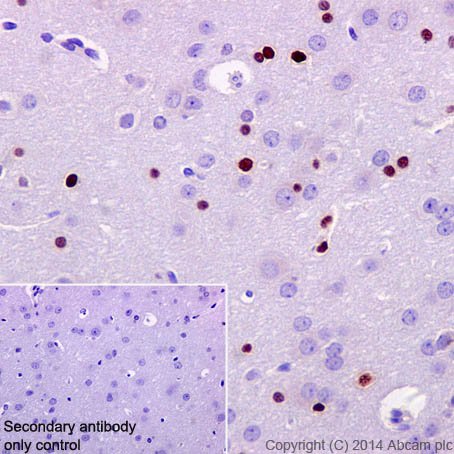
Immunohistochemical staining of paraffin embedded rat cerebral cortex with purified ab109186 at a working dilution of 1/100. The secondary antibody used is ab97051, a HRP-conjugated goat anti-rabbit IgG (H+L), at a dilution of 1/500. The sample is counter-stained with hematoxylin. Antigen retrieval was perfomed using Tris-EDTA buffer, pH 9.0. PBS was used instead of the primary antibody as the negative control, and is shown in the inset.
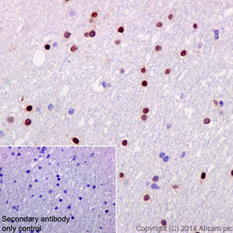
Immunohistochemical staining of paraffin embedded human cerebral cortex with purified ab109186 at a working dilution of 1/100. The secondary antibody used is ab97051, a HRP-conjugated goat anti-rabbit IgG (H+L), at a dilution of 1/500. The sample is counter-stained with hematoxylin. Antigen retrieval was perfomed using Tris-EDTA buffer, pH 9.0. PBS was used instead of the primary antibody as the negative control, and is shown in the inset.
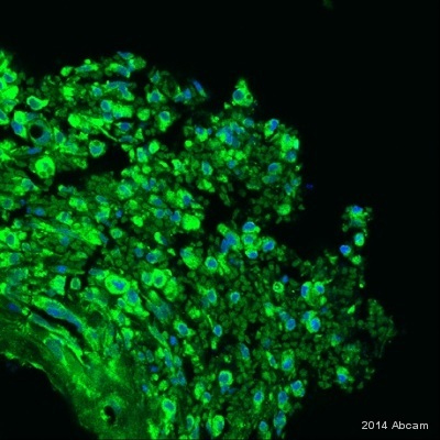
Unpurified ab109186 staining Olig2 in rat brain tissue sections by immunohistochemistry (IHC-Fr - frozen sections). Tissue was fixed with paraformaldehyde, permeabilized with 0.3% Triton X-100 in 0.1M PBS and blocked with 10% serum for 1 hour at 24°C. Samples were incubated with primary antibody (1/200 in PBS + 10% donkey serum) for 4 hours at 24°C. An Alexa Fluor® 488-conjugated anti-rabbit IgG polyclonal (1/1000) was used as the secondary antibody. Nuclei stained with Hoechst (blue).See Abreview
![Anti-Olig2 antibody [EPR2673] (ab109186) at 1/1000 dilution (unpurified) + Oligodendroglioma lysate at 10 µgPerformed under reducing conditions.](http://www.bioprodhub.com/system/product_images/ab_products/2/sub_4/3780_Olig2-Primary-antibodies-ab109186-1.jpg)
Anti-Olig2 antibody [EPR2673] (ab109186) at 1/1000 dilution (unpurified) + Oligodendroglioma lysate at 10 µgPerformed under reducing conditions.
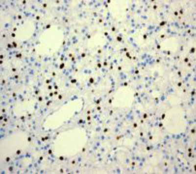
Immunohistochemical staining of Olig2 in human glioma tissue with ab109186 at a dilution of 1/100.
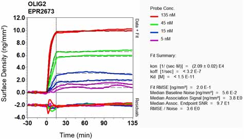
Equilibrium disassociation constant (KD)Learn more about KD Click here to learn more about KD
![All lanes : Anti-Olig2 antibody [EPR2673] (ab109186) at 1/2000 dilution (purified)Lane 1 : Mouse brain lysateLane 2 : Mouse brain lysateLysates/proteins at 20 µg per lane.SecondaryHRP goat anti-rabbit (H+L) at 1/1000 dilution](http://www.bioprodhub.com/system/product_images/ab_products/2/sub_4/3774_ab109186-7-ab109186WB3.jpg)
![Anti-Olig2 antibody [EPR2673] (ab109186) at 1/10000 dilution (purified) + Human oligodendroglioma lysate at 10 µgSecondaryHRP goat anti-rabbit (H+L) at 1/1000 dilution](http://www.bioprodhub.com/system/product_images/ab_products/2/sub_4/3775_ab109186-6-ab109186WB2.jpg)
![Anti-Olig2 antibody [EPR2673] (ab109186) at 1/2000 dilution (purified) + Human fetal brain tissue lysate at 20 µgSecondaryHRP goat anti-rabbit (H+L) at 1/1000 dilution](http://www.bioprodhub.com/system/product_images/ab_products/2/sub_4/3776_ab109186-5-ab109186WB1.jpg)


![Anti-Olig2 antibody [EPR2673] (ab109186) at 1/1000 dilution (unpurified) + Oligodendroglioma lysate at 10 µgPerformed under reducing conditions.](http://www.bioprodhub.com/system/product_images/ab_products/2/sub_4/3780_Olig2-Primary-antibodies-ab109186-1.jpg)

