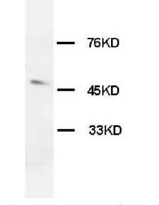
Anti-Orexin Receptor 1 antibody (ab68718) at 2 µg/ml + rat brain tissue lysate
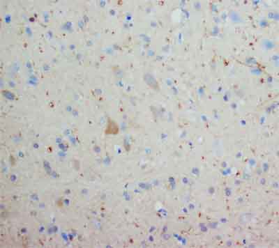
ab68718 staining Orexin Receptor 1 in rat brain tissue by Immunohistochemistry (formalin fixed, paraffin embedded tissue section). Primary antibody used at 2µg/ml.
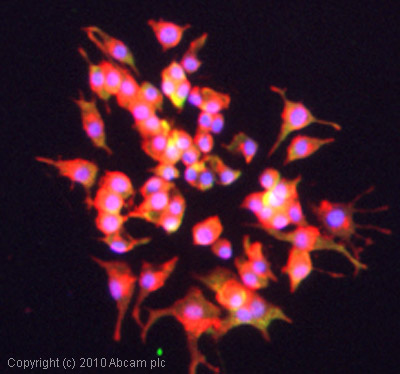
ICC/IF image of ab68718 stained PC12 cells. The cells were 100% methanol fixed (5 min) and then incubated in 1%BSA / 10% normal goat serum / 0.3M glycine in 0.1% PBS-Tween for 1h to permeabilise the cells and block non-specific protein-protein interactions. The cells were then incubated with the antibody (ab68718, 5µg/ml) overnight at +4°C. The secondary antibody (green) was Alexa Fluor® 488 goat anti-rabbit IgG (H+L) used at a 1/1000 dilution for 1h. Alexa Fluor® 594 WGA was used to label plasma membranes (red) at a 1/200 dilution for 1h. DAPI was used to stain the cell nuclei (blue) at a concentration of 1.43µM.
![Orexin Receptor 1 was immunoprecipitated using 0.5mg Rat Brain tissue lysate, 5µg of Rabbit polyclonal to Orexin Receptor 1 and 50µl of protein G magnetic beads (+). No antibody was added to the control (-). The antibody was incubated under agitation with Protein G beads for 10min, Rat Brain tissue lysate lysate diluted in RIPA buffer was added to each sample and incubated for a further 10min under agitation.Proteins were eluted by addition of 40µl SDS loading buffer and incubated for 10min at 70oC; 10µl of each sample was separated on a SDS PAGE gel, transferred to a nitrocellulose membrane, blocked with 5% BSA and probed with ab68718.Secondary: Mouse monoclonal [SB62a] Secondary Antibody to Rabbit IgG light chain (HRP) (ab99697).Band: 52kDa; Orexin Receptor 1](http://www.bioprodhub.com/system/product_images/ab_products/2/sub_4/4623_Orexin-Receptor-1-Primary-antibodies-ab68718-4.jpg)
Orexin Receptor 1 was immunoprecipitated using 0.5mg Rat Brain tissue lysate, 5µg of Rabbit polyclonal to Orexin Receptor 1 and 50µl of protein G magnetic beads (+). No antibody was added to the control (-). The antibody was incubated under agitation with Protein G beads for 10min, Rat Brain tissue lysate lysate diluted in RIPA buffer was added to each sample and incubated for a further 10min under agitation.Proteins were eluted by addition of 40µl SDS loading buffer and incubated for 10min at 70oC; 10µl of each sample was separated on a SDS PAGE gel, transferred to a nitrocellulose membrane, blocked with 5% BSA and probed with ab68718.Secondary: Mouse monoclonal [SB62a] Secondary Antibody to Rabbit IgG light chain (HRP) (ab99697).Band: 52kDa; Orexin Receptor 1