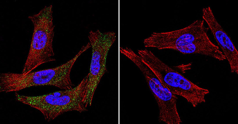
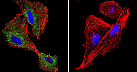
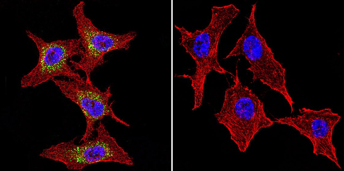
Anti-P cadherin antibody [6A9]
| Name | Anti-P cadherin antibody [6A9] |
|---|---|
| Supplier | Abcam |
| Catalog | ab19350 |
| Prices | $373.00 |
| Sizes | 100 µg |
| Host | Mouse |
| Clonality | Monoclonal |
| Isotype | IgG1 |
| Clone | 6A9 |
| Applications | ICC/IF ICC/IF WB IP B/N |
| Species Reactivities | Human |
| Antigen | Full length protein corresponding to P cadherin |
| Description | Mouse Monoclonal |
| Gene | CDH3 |
| Conjugate | Unconjugated |
| Supplier Page | Shop |
Product images
Product References
Mimicking normal tissue architecture and perturbation in cancer with engineered - Mimicking normal tissue architecture and perturbation in cancer with engineered
Gautrot JE, Wang C, Liu X, Goldie SJ, Trappmann B, Huck WT, Watt FM. Biomaterials. 2012 Jul;33(21):5221-9.
Cross-talk between adherens junctions and desmosomes depends on plakoglobin. - Cross-talk between adherens junctions and desmosomes depends on plakoglobin.
Lewis JE, Wahl JK 3rd, Sass KM, Jensen PJ, Johnson KR, Wheelock MJ. J Cell Biol. 1997 Feb 24;136(4):919-34.
![All lanes : Anti-P cadherin antibody [6A9] (ab19350) at 1/500 dilutionLane 1 : 293T cell lysateLane 2 : A431 cell lysateLane 3 : Mouse brain tissue lysateLysates/proteins at 25 µg per lane.SecondaryHRP-conjugated anti-mouse IgG](http://www.bioprodhub.com/system/product_images/ab_products/2/sub_4/5159_ab19350-241916-ab19350wb.jpg)


