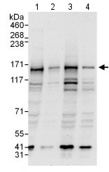
All lanes : Anti-P Glycoprotein antibody (ab129450) at 0.4 µg/mlLane 1 : HeLa whole cell lysate at 50 µgLane 2 : HeLa whole cell lysate at 15 µgLane 3 : 293T whole cell lysate at 50 µgLane 4 : Jurkat whole cell lysate at 50 µgdeveloped using the ECL technique

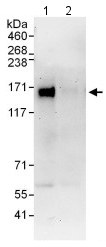
Detection of P Glycoprotein by Western Blot of Immunprecipitate. Lane 1: ab129450 at 1µg/ml staining P Glycoprotein in HeLa whole cell lysate immunoprecipitated using ab129450 at 6µg/mg lysate (1 mg/IP; 20% of IP loaded/lane). Lane 2: Control IgG Detection: Chemiluminescence with exposure time of 30 seconds.

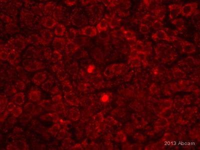
ICC/IF image of ab129450 stained human hepatocyte-like cells. The cells were fixed in methanol and then incubated in 10% BSA for 1 hour to block non-specific protein-protein interactions. The cells were then incubated with the antibody (ab129450, 1/200) overnight at +4°C. The secondary antibody (red) was Alexa Fluor® 568 goat anti-rabbit used at a 1/400 dilution.See Abreview


