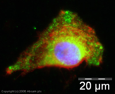
Anti-p53 (phospho S315) antibody
| Name | Anti-p53 (phospho S315) antibody |
|---|---|
| Supplier | Abcam |
| Catalog | ab1647 |
| Prices | $370.00 |
| Sizes | 50 µg |
| Host | Rabbit |
| Clonality | Polyclonal |
| Isotype | IgG |
| Applications | IHC-P WB ICC/IF ICC/IF |
| Species Reactivities | Human |
| Antigen | Synthetic peptide derived from within residues 300 to the C-terminus of Human p53 |
| Blocking Peptide | Human p53 (phospho S315) peptide |
| Description | Rabbit Polyclonal |
| Gene | TP53 |
| Conjugate | Unconjugated |
| Supplier Page | Shop |
Product images
Product References
Diabetes-induced oxidative dna damage alters p53-p21CIP1/Waf1 signaling in the - Diabetes-induced oxidative dna damage alters p53-p21CIP1/Waf1 signaling in the
Kilarkaje N, Al-Bader MM. Reprod Sci. 2015 Jan;22(1):102-12.
Choice of fixative is crucial to successful immunohistochemical detection of - Choice of fixative is crucial to successful immunohistochemical detection of
Burns JA, Li Y, Cheney CA, Ou Y, Franlin-Pfeifer LL, Kuklin N, Zhang ZQ. J Histochem Cytochem. 2009 Mar;57(3):257-64.


