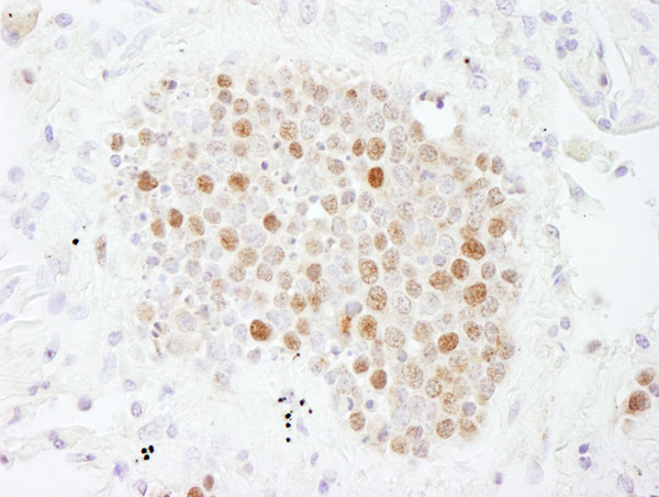
Immunohistochemistry (Formalin/PFA-fixed paraffin-embedded sections) analysis of human small cell lung cancer tissue labelling p60 CAF1 with ab7252 at 1/200 (1µg/ml). Detection: DAB.

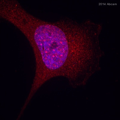
ab72520 staining p60 CAF1 in human PC3 prostate cancer cells by ICC/IF (Immunocytochemistry/immunofluorescence). Cells were fixed with paraformaldehyde, permeabilized with 0.1% Triton X-100 pH 7.4 for 5 minutes at room temperature and blocked with 5% BSA for 20 minutes at room temperature. Samples were incubated with primary antibody (1/100 in PBS) for 1 hour. A CF568-conjugated donkey anti-rabbit IgG polyclonal (1/500) was used as the secondary antibody.See Abreview

All lanes : Anti-p60 CAF1 antibody (ab72520) at 0.04 µg/mlLane 1 : Whole cell lysate from Hela cells at 50 µgLane 2 : Whole cell lysate from Hela cells at 15 µgLane 3 : Whole cell lysate from Hela cells at 5 µgLane 4 : Whole cell lysate from 293T cells at 50 µgdeveloped using the ECL technique

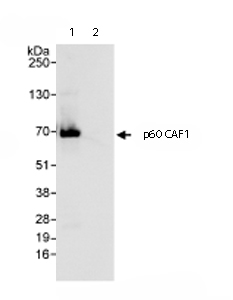
Immunoprecipitation/ Western Blot of p60 CAF1. Lane 1: ab72520 at 3µg/mg whole cell lysate. Lane 2: Control IgG. ab72520 at 1µg/ml for WB. Whole cell lysate from Hela cells at 1mg for IP, 20% of IP loaded. Chemiluminescence with an exposure time of 3 seconds.


