![Anti-p63 antibody [Y289] (ab32353) at 1/4000 dilution (unpurified) + A431 cell lysate at 10 µgSecondaryPeroxidase-conjugated goat anti-rabbit IgG (H+L) at 1/1000 dilution](http://www.bioprodhub.com/system/product_images/ab_products/2/sub_4/6092_ab32353-241828-ab32353upwb.jpg)
Anti-p63 antibody [Y289] (ab32353) at 1/4000 dilution (unpurified) + A431 cell lysate at 10 µgSecondaryPeroxidase-conjugated goat anti-rabbit IgG (H+L) at 1/1000 dilution
![Anti-p63 antibody [Y289] (ab32353) at 1/10000 dilution (purified) + A431 cell lysate at 10 µgSecondaryPeroxidase-conjugated goat anti-rabbit IgG (H+L) at 1/1000 dilution](http://www.bioprodhub.com/system/product_images/ab_products/2/sub_4/6093_ab32353-241829-ab32353pwb.jpg)
Anti-p63 antibody [Y289] (ab32353) at 1/10000 dilution (purified) + A431 cell lysate at 10 µgSecondaryPeroxidase-conjugated goat anti-rabbit IgG (H+L) at 1/1000 dilution
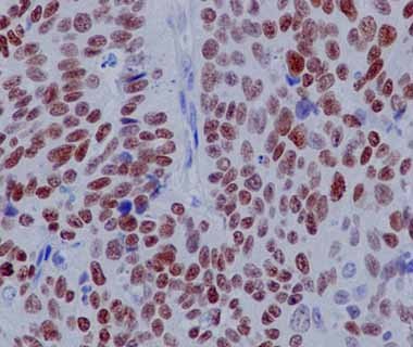
Immunohistochemistry (Formalin/PFA-fixed paraffin-embedded sections) analysis of human hepatocellular skin tissue labelling p63 with purified ab32353 at 1/50. Heat mediated antigen retrieval was performed using Tris/EDTA buffer pH 9. A prediluted HRP-polymer conjugated anti-rabbit IgG was used as the secondary antibody. Counterstained with Hematoxylin.
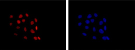
Immunocytochemsitry/Immunofluorescence analysis of A431 cells labelling p63 (red) with purified ab32353 at 1/200. Cells were fixed with 4% paraformaldehyde. An Alexa Fluor® 555-conjugated goat anti-rabbit IgG (1/200) was used as the secondary antibody. Counterstained with DAPI (blue).
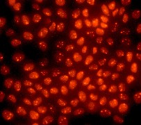
Immunofluorescent staining of A431 cells using ab32353 at 1/250 dilution.