TP63

Gene Symbol TP63
Entrez Gene 8626
Alt Symbol AIS, B(p51A), B(p51B), EEC3, KET, LMS, NBP, OFC8, RHS, SHFM4, TP53CP, TP53L, TP73L, p40, p51, p53CP, p63, p73H, p73L
Species Human
Gene Type protein-coding
Description tumor protein p63
Other Description CUSP|amplified in squamous cell carcinoma|chronic ulcerative stomatitis protein|keratinocyte transcription factor KET|transformation-related protein 63|tumor protein 63|tumor protein p53-competing protein|tumor protein p63 deltaN isoform delta
Swissprots Q6VFJ3 Q9H3D3 Q9UP74 Q9UP26 Q9P1B7 O75080 Q9UE10 O75922 Q6VEG3 Q9H3P8 Q6VEG2 Q9H3D4 O76078 Q9UP27 Q6VH20 Q6VFJ1 Q9NPH7 Q7LDI4 Q9H3D2 Q6VFJ2 Q6VEG4 Q9UBV9 Q9P1B5 Q7LDI3 Q96KR0 Q9UP28 Q9P1B6 Q9P1B4 O75195 Q7LDI5
Accessions AAF43486 AAF43487 AAF43488 AAF43489 AAF43490 AAF43491 AAF43492 AAF43493 AAG45607 AAG45608 AAG45609 AAG45610 AAG45611 AAG45612 AAQ63448 AAQ63449 AAQ63450 AAQ63451 AAQ63452 AAQ63453 AAQ63454 CDI30201 EAW78112 EAW78113 EAW78114 EAW78115 EAW78116 EAW78117 EAW78118 EAW78119 EAW78120 Q9H3D4 AB010153 BAA32433 AB016072 BAA32592 AB016073 BAA32593 AB042841 BAB20591 AF075428 AAC62633 AF075429 AAC62634 AF075430 AAC62635 AF075431 AAC62636 AF075432 AAC62637 AF075433 AAC62638 AF091627 AAC43038 AF116771 AAF61624 AJ315499 CAC48053 AK304127 BAH14112 BC039815 AAH39815 CB241431 EU446669 ABZ92198 GQ202689 ACV53609 GQ202690 ACV53610 Y16961 CAA76562 XM_005247843 XP_005247900 XM_005247844 XP_005247901 XM_005247846 XP_005247903 XM_011513251 XP_011511553 XM_011513252 XP_011511554 XM_011513253 XP_011511555 NM_001114978 NP_001108450 NM_001114979 NP_001108451 NM_001114980 NP_001108452 NM_001114981 NP_001108453 NM_001114982 NP_001108454 NM_003722 NP_003713
Function Acts as a sequence specific DNA binding transcriptional activator or repressor. The isoforms contain a varying set of transactivation and auto-regulating transactivation inhibiting domains thus showing an isoform specific activity. Isoform 2 activates RIPK4 transcription. May be required in conjunction with TP73/p73 for initiation of p53/TP53 dependent apoptosis in response to genotoxic insults and the presence of activated oncogenes. Involved in Notch signaling by probably inducing JAG1 and JAG2. Plays a role in the regulation of epithelial morphogenesis. The ratio of DeltaN-type and TA*-type isoforms may govern the maintenance of epithelial stem cell compartments and regulate the initiation of epithelial stratification from the undifferentiated embryonal ectoderm. Required for limb formation from the apical ectodermal ridge. Activates transcription of the p21 promoter. {ECO:0000269|PubMed:11641404, ECO:0000269|PubMed:12374749, ECO:0000269|PubMed:12446779, ECO:0000269|PubMed:12446784,
Subcellular Location Nucleus {ECO:0000269|PubMed:12446779, ECO:0000269|PubMed:20123734}.
Tissue Specificity Widely expressed, notably in heart, kidney, placenta, prostate, skeletal muscle, testis and thymus, although the precise isoform varies according to tissue type. Progenitor cell layers of skin, breast, eye and prostate express high levels of DeltaN-type isoforms. Isoform 10 is predominantly expressed in skin squamous cell carcinomas, but not in normal skin tissues. {ECO:0000269|PubMed:11248048, ECO:0000269|PubMed:9774969}.
Top Pathways MicroRNAs in cancer

Phospho-p63 (Ser160/162) Antibody - 4981 from Cell Signaling Technology

Prices $287.00
Sizes 100 µl (10 western blots)
Host Rabbit
Clonality Polyclonal
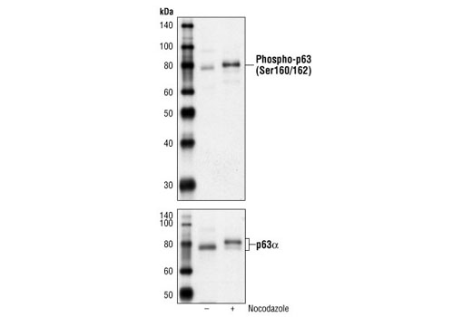
Western blot analysis of extracts from untreated or nocodazole-treated (50 ng/ml, 24 h) ME-180 cells using Phospho-p63 (Ser160/162) Antibody (upper) or p63alpha Antibody #4892 (lower).

p63-α (D2K8X) XP ® Rabbit mAb - 13109 from Cell Signaling Technology

Prices $109.00, $277.00
Sizes 20 µl (2 western blots), 100 µl (10 western blots)
Host Rabbit
Clonality Monoclonal
Clone D2K8X
Western blot analysis of extracts from ME-180 (+), HaCaT (+), and MCF7 (-) cells using p63-α (D2K8X) XP ® Rabbit mAb (upper) or β-Actin (D6A8) Rabbit mAb #8457 (lower).

p63-α Antibody - 4892 from Cell Signaling Technology

Prices $246.00
Sizes 100 µl (10 western blots)
Host Rabbit
Clonality Polyclonal
Western blot analysis of extracts from ME-180 cells, using p63α Antibody.

Phospho-p63 (Ser455) Antibody - 4985 from Cell Signaling Technology

Host Rabbit
Clonality Polyclonal
Western blot analysis of extracts from untreated or nocodazole-treated (50 ng/ml, 24 h) ME-180 cells, using Phospho-p63 (Ser455) Antibody (upper) or p63alpha Antibody #4892 (lower).

Phospho-p63 (Ser395) Antibody - 4984 from Cell Signaling Technology

Host Rabbit
Clonality Polyclonal

Anti-p51A antibody - ab2506 from Abcam

Host Rabbit
Clonality Polyclonal

Anti-p51B antibody - ab2505 from Abcam

Host Rabbit
Clonality Polyclonal

Anti-p63 (phospho S395) antibody - ab92581 from Abcam

Host Rabbit
Clonality Polyclonal
All lanes : Anti-p63 (phospho S395) antibody (ab92581) at 1/500 dilutionLane 1 : Extracts from HepG2 cells treated with nocodazole (1 µg/ml, 18 hours) Lane 2 : Extracts from A549 cells treated with nocodazole (1 µg/ml, 18 hours) Lane 3 : Extracts from LOVO cells treated with nocodazole (1 µg/ml, 18 hours) Lane 4 : Extracts from HepG2 cells treated with nocodazole (1 µg/ml, 18 hours) with immunizing peptide at 10 µgLysates/proteins at 30 µg per lane.

Anti-p63 (phospho S455) antibody - ab79200 from Abcam

Host Rabbit
Clonality Polyclonal
All lanes : Anti-p63 (phospho S455) antibody (ab79200) at 1/500 dilutionLane 1 : Extracts from HeLa cells treated with TNF (2500U/ml, 30mins)Lane 2 : Extracts from HeLa cells treated with TNF (2500U/ml, 30mins) with immunising phosphopeptide

Anti-p63 (phospho S455) antibody - ab63362 from Abcam

Host Rabbit
Clonality Polyclonal
Immunohistochemistry analysis of p63 in human breast carcinoma tissue (paraffin section) using ab63362 at 1/50 dilution in the presence and absence of the immunizing phosphopeptide.

Anti-p63 antibody - ab53039 from Abcam

Host Rabbit
Clonality Polyclonal
All lanes : Anti-p63 antibody (ab53039) at 1/500 dilutionLane 1 : Cos7 cell extractLane 2 : Cos7 cell extract with peptide at 1 µg/ml

Anti-p63 antibody - ab63881 from Abcam

Host Rabbit
Clonality Polyclonal
ICC/IF image of ab63881 stained A431 cells. The cells were 4% Formaldehyde fixed (10 min) and then incubated in 1%BSA / 10% normal Goat serum / 0.3M glycine in 0.1% PBS-Tween for 1h to permeabilise the cells and block non-specific protein-protein interactions. The cells were then incubated with the antibody (ab63881, 5µg/ml) overnight at +4°C. The secondary antibody (green) was ab96899, DyLight® 488 Goat anti-Rabbit IgG (H+L) used at a 1/250 dilution for 1h. Alexa Fluor® 594 WGA was used to label plasma membranes (red) at a 1/200 dilution for 1h. DAPI was used to stain the cell nuclei (blue) at a concentration of 1.43µM.

Anti-p63 antibody - ab97865 from Abcam

Host Rabbit
Clonality Polyclonal
Anti-p63 antibody (ab97865) at 1/500 dilution + A431 whole cell lysate at 30 µg

Anti-p63 antibody - ab97866 from Abcam

Host Rabbit
Clonality Polyclonal
Anti-p63 antibody (ab97866) at 1/1000 dilution + Raji whole cell lysate at 30 µg

Anti-p63 antibody [4E5] - ab110038 from Abcam

Host Mouse
Clonality Monoclonal
Clone 4E5
All lanes : Anti-p63 antibody [4E5] (ab110038) at 1/500 dilutionLane 1 : A431 cell lysate.Lane 2 : HeLa cell lysate.Lane 3 : Jurkat cell lysate.Lane 4 : THP1 cell lysate.Lane 5 : NIH 3T3 cell lysate.Lane 6 : COS7 cell lysate.Lane 7 : PC12 cell lysate.

Anti-p63 antibody [87C553] - ab11995 from Abcam

Host Mouse
Clonality Monoclonal
Clone 87C553

Anti-p63 antibody [C24-I] - N-terminal - ab179874 from Abcam

Host Rabbit
Clonality Monoclonal
Clone C24-I
All lanes : Anti-p63 antibody [C24-I] - N-terminal (ab179874) at 1/1000 dilutionLane 1 : Recombinant tagged Human AMACR + p63 (aa 1 – 680) at 0.1 µgLane 2 : Recombinant tagged Human AMACR + p63 (aa 1 – 680) at 0.2 µgLane 3 : Recombinant tagged Human AMACR + p63 (aa 1 – 680) at 0.5 µg

Anti-p63 antibody [EPR5701] - ab124762 from Abcam

Host Rabbit
Clonality Monoclonal
Clone EPR5701
Immunofluorescent staining of A431 cells (fixed with 4% PFA and permeablized with TritonX 100) with purified ab124762 at a dilution of 1/60. An Alexa Fluor® 488 goat anti-rabbit antibody was used as the secondary at a dilution of 1/200. The panel on the right shows the DAPI counter-staining.

Anti-p63 antibody [EPR5701] (DyLight® 488) - ab139929 from Abcam

Host Rabbit
Clonality Monoclonal
Clone EPR5701
Immunofluorescence analysis of A431 cells labelling p63 with ab139929 at 1/100 dilution (green); Dapi (blue).

Anti-p63 antibody [I27-I] - ab169707 from Abcam

Host Rabbit
Clonality Monoclonal
Clone I27-I
Immunohistochemical analysis of Human normal hair follicle tissue labeling p63 with ab169707 at 1/100.

Anti-p63 antibody [V22-E] - ab191893 from Abcam

Host Rabbit
Clonality Monoclonal
Clone V22-E
Nuclear expression of p63 visualized with ab191893 a rabbit monclonal anti-p63 [V22-E]  in basal cell carcinoma of the skin.

Anti-p63 antibody [Y289] - ab32353 from Abcam

Host Rabbit
Clonality Monoclonal
Clone Y289
Anti-p63 antibody [Y289] (ab32353) at 1/4000 dilution (unpurified) + A431 cell lysate at 10 µgSecondaryPeroxidase-conjugated goat anti-rabbit IgG (H+L) at 1/1000 dilution

p63/TP73L Antibody [Unconjugated] - AF1916 from Novus Biologicals

Prices $99.00, $359.00
Sizes 25 µg, 100 µg
Host Goat
Clonality Polyclonal
p63/TP73L was detected in immersion fixed paraffin-embedded sections of human breast using Goat Anti-Human p63/TP73L Antigen Affinity-purified Polyclonal Antibody (Catalog # AF1916) at 15 µg/mL overnight at 4 °C. Tissue was stained using the Anti-Goat HRP-DAB Cell & Tissue Staining Kit (brown; Catalog # CTS008) and counterstained with hematoxylin (blue). Specific staining was localized to nuclei. View our protocol for Chromogenic IHC Staining of Paraffin-embedded Tissue Sections.

p63/TP73L Antibody (241631) - MAB1916 from Novus Biologicals

Host Mouse
Clonality Monoclonal
Clone 241631

p63/TP73L Antibody [Biotin] - BAF1916 from Novus Biologicals

Prices $390.00
Sizes 50 µg
Host Goat
Clonality Polyclonal
p63/TP73L was detected in immersion fixed SCC-25 human tongue carcinoma cell line using Goat Anti-Human p63/TP73L Biotinylated Antigen Affinity-purified Polyclonal Antibody (Catalog # BAF1916) at 10 µg/mL for 3 hours at room temperature. Cells were stained using the NorthernLights™ 557-conjugated Streptavidin (yellow; Catalog # NL999) and counterstained with DAPI (blue). Specific staining was localized to nuclei. View our protocol for Fluorescent ICC Staining of Cells on Coverslips.
Search more