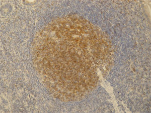
Anti-PAG antibody [PAG-C1]
| Name | Anti-PAG antibody [PAG-C1] |
|---|---|
| Supplier | Abcam |
| Catalog | ab4206 |
| Prices | $385.00 |
| Sizes | 100 µg |
| Host | Mouse |
| Clonality | Monoclonal |
| Isotype | IgG2b |
| Clone | PAG-C1 |
| Applications | IHC-P IP WB FC |
| Species Reactivities | Mouse, Rat, Bovine, Human |
| Antigen | Synthetic C-teminal peptide (last 15 amino acids) (Human) conjugated to KLH |
| Description | Mouse Monoclonal |
| Gene | PAG1 |
| Conjugate | Unconjugated |
| Supplier Page | Shop |
Product images
Product References
Constitutive exclusion of Csk from Hck-positive membrane microdomains permits Src - Constitutive exclusion of Csk from Hck-positive membrane microdomains permits Src
Baumgartner M, Angelisova P, Setterblad N, Mooney N, Werling D, Horejsi V, Langsley G. Blood. 2003 Mar 1;101(5):1874-81. Epub 2002 Oct 31.
Combined spatial and enzymatic regulation of Csk by cAMP and protein kinase a - Combined spatial and enzymatic regulation of Csk by cAMP and protein kinase a
Vang T, Abrahamsen H, Myklebust S, Horejsi V, Tasken K. J Biol Chem. 2003 May 16;278(20):17597-600. Epub 2003 Mar 28.
![Overlay histogram showing Jurkat cells stained with ab4206 (red line). The cells were fixed with 4% paraformaldehyde (10 min) and then permeabilized with 0.1% PBS-Tween for 20 min. The cells were then incubated in 1x PBS / 10% normal goat serum / 0.3M glycine to block non-specific protein-protein interactions followed by the antibody (ab4206, 1μg/1x106 cells) for 30 min at 22°C. The secondary antibody used was Alexa Fluor® 488 goat anti-mouse IgG (H+L) (ab150113) at 1/2000 dilution for 30 min at 22°C. Isotype control antibody (black line) was mouse IgG2b [PLPV219] (ab91366, 1μg/1x106 cells) used under the same conditions. Unlabelled sample (blue line) was also used as a control. Acquisition of >5,000 events were collected using a 20mW Argon ion laser (488nm) and 525/30 bandpass filter. This antibody gave a positive signal in Jurkat cells fixed with 80% methanol (5 min)/permeabilized with 0.1% PBS-Tween for 20 min used under the same conditions.](http://www.bioprodhub.com/system/product_images/ab_products/2/sub_4/6539_ab4206-3-ab4206FC.jpg)

