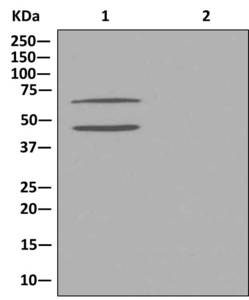
Western blot analysis on immunoprecipitation pellet from (1) HeLa cell lysate or (2) 1XPBS (negative control) using ab181359 at 1/10 dilution, and HRP-conjugated anti-rabbit IgG preferentially detecting the non-reduced form of rabbit IgG
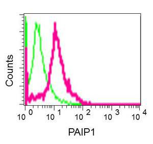
Flow cytometric analysis of permeabilized 293 cells using ab181359 at 1/10 dilution (red) or a rabbit IgG (negative) (green).
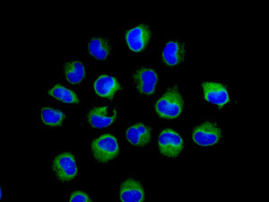
Immunofluorescence analysis of HeLa cells labeling PAIP1 with ab181359 at 1/50 dilution (green). DAPI nuclear staining (blue).
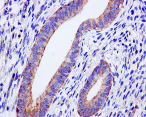
Immunohistochemical analysis of paraffin-embedded Human uterus tissue labeling PAIP1 with ab181359 at 1/50 dilution.
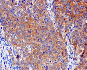
Immunohistochemical analysis of paraffin-embedded Human gastric carcinoma tissue labeling PAIP1 with ab181359 at 1/50 dilution.
![All lanes : Anti-PAIP1 antibody [EPR13259] (ab181359) at 1/1000 dilutionLane 1 : HepG2 cell lysatesLane 2 : HeLa cell lysatesLane 3 : 293T cell lysatesLysates/proteins at 10 µg per lane.](http://www.bioprodhub.com/system/product_images/ab_products/2/sub_4/6649_ab181359-210434-Image1.jpg)
All lanes : Anti-PAIP1 antibody [EPR13259] (ab181359) at 1/1000 dilutionLane 1 : HepG2 cell lysatesLane 2 : HeLa cell lysatesLane 3 : 293T cell lysatesLysates/proteins at 10 µg per lane.


![All lanes : Anti-PAIP1 antibody [EPR13259] (ab181359) at 1/1000 dilutionLane 1 : HepG2 cell lysatesLane 2 : HeLa cell lysatesLane 3 : 293T cell lysatesLysates/proteins at 10 µg per lane.](http://www.bioprodhub.com/system/product_images/ab_products/2/sub_4/6649_ab181359-210434-Image1.jpg)