ab75150 staining Parathyroid Hormone Receptor 1 in Lizard regenerated tail (Anolis sagrei) tissue sections by Immunohistochemistry (IHC-P - paraformaldehyde-fixed, paraffin-embedded sections). Tissue was fixed with paraformaldehyde and blocked with 1% serum for 45 minutes at 22°C; antigen retrieval was by heat mediation in sodium citrate buffer, pH 6.0. Samples were incubated with primary antibody (1/200 in 1% horse serum) for 16 hours at 4°C. A Biotin-conjugated horse anti-rabbit IgG polyclonal (1/50) was used as the secondary antibody.Image is of a regenerated lizard tail cartilage tube, at a proximal region that has begun to ossify. Positive staining was observed in structures resembling embryonic bone collar and early hypertrophic chondrocytes.See Abreview

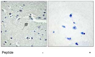
Immunohistochemistry analysis of Parathyroid Hormone Receptor 1 in paraffin-embedded human brain tissue using ab75150, at 1/50 dilution, in the presence (right panel) or absence (left panel) of immunising peptide.

All lanes : Anti-Parathyroid Hormone Receptor 1 antibody (ab75150) at 1/500 dilutionLane 1 : Extracts from Jurkat cellsLane 2 : Extracts from Jurkat cells with immunising peptide at 5 µgLysates/proteins at 5 µg per lane.

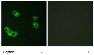
Immunofluorescence analysis of Parathyroid Hormone Receptor 1 in MCF7 cells using ab75150, at 1/500 dilution, in the presence (right panel) or absence (left panel) of immunising peptide.


