Anti-PARG antibody
| Name | Anti-PARG antibody |
|---|---|
| Supplier | Abcam |
| Catalog | ab16060 |
| Prices | $400.00 |
| Sizes | 100 µg |
| Host | Rabbit |
| Clonality | Polyclonal |
| Isotype | IgG |
| Applications | IHC-P ICC/IF ICC/IF IHC-F WB |
| Species Reactivities | Mouse, Human |
| Antigen | Synthetic peptide conjugated to KLH derived from within residues 100 - 200 of Human PARG |
| Description | Rabbit Polyclonal |
| Gene | PARG |
| Conjugate | Unconjugated |
| Supplier Page | Shop |
Product images
 Lanes 1 - 5 : Anti-PARG antibody (ab16060)Lanes 6 - 10 : Anti-PARG antibody (ab16060) at 0.1 µgLane 1 : Hela nuclear Lane 2 : Hela WC Lane 3 : A431Lane 4 : Mouse Brain Lane 5 : Mouse Liver Lane 6 : Hela nuclear with Human PARG peptide (ab16386) at 1 µg/mlLane 7 : Hela WC with Human PARG peptide (ab16386) at 1 µg/mlLane 8 : A431 with Human PARG peptide (ab16386) at 1 µg/mlLane 9 : Mouse Brain with Human PARG peptide (ab16386) at 1 µg/mlLane 10 : Mouse Liver with Human PARG peptide (ab16386) at 1 µg/mlLysates/proteins at 20 µg per lane.SecondaryAlexa Fluor Goat polyclonal to Rabbit IgG (700) (1/10,000)
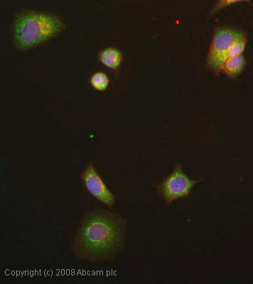 ICC/IF image of ab16060 stained human MCF7 cells. The cells were 4% PFA fixed (10 min), permabilised in 0.1% PBS-Tween (20 min) and incubated with the antibody (ab16060, 5µg/ml) for 1h at room temperature. 1%BSA / 10% normal goat serum / 0.3M glycine was used to block non-specific protein-protein interactions. The secondary antibody (green) was Alexa Fluor® 488 goat anti-rabbit IgG (H+L) used at a 1/1000 dilution for 1h. Alexa Fluor® 594 WGA was used to label plasma membranes (red). DAPI was used to stain the cell nuclei (blue). This antibody also gave a positive IF result in HeLa, HEK293 and HepG2 cells.
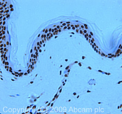 IHC image of PARG staining in human skin FFPE section, performed on a Leica BondTM system using the standard protocol F. The section was pre-treated using heat mediated antigen retrieval with sodium citrate buffer (pH6, epitope retrieval solution 1) for 20 mins. The section was then incubated with ab16060, 1µg/ml, for 15 mins at room temperature and detected using an HRP conjugated compact polymer system. DAB was used as the chromogen. The section was then counterstained with haematoxylin and mounted with DPX.
Product References
Knockout of PARG110 confers resistance to cGMP-induced toxicity in mammalian - Knockout of PARG110 confers resistance to cGMP-induced toxicity in mammalian
Sahaboglu A, Tanimoto N, Bolz S, Garrido MG, Ueffing M, Seeliger MW, Lowenheim H, Ekstrom P, Paquet-Durand F. Cell Death Dis. 2014 May 22;5:e1234.
Mutational analysis of the poly(ADP-ribosyl)ation sites of the transcription - Mutational analysis of the poly(ADP-ribosyl)ation sites of the transcription
Farrar D, Rai S, Chernukhin I, Jagodic M, Ito Y, Yammine S, Ohlsson R, Murrell A, Klenova E. Mol Cell Biol. 2010 Mar;30(5):1199-216.
Poly(ADP-ribose) polymerase 1 accelerates single-strand break repair in concert - Poly(ADP-ribose) polymerase 1 accelerates single-strand break repair in concert
Fisher AE, Hochegger H, Takeda S, Caldecott KW. Mol Cell Biol. 2007 Aug;27(15):5597-605. Epub 2007 Jun 4.