Anti-PARP4 antibody
| Name | Anti-PARP4 antibody |
|---|---|
| Supplier | Abcam |
| Catalog | ab86066 |
| Prices | $370.00 |
| Sizes | 100 µl |
| Host | Rabbit |
| Clonality | Polyclonal |
| Isotype | IgG |
| Applications | WB IP IHC-P |
| Species Reactivities | Human, Chimpanzee, Monkey, Orangutan |
| Antigen | Synthetic peptide corresponding to a region within N terminal amino acids 1674-1724 ( EWVRRTEGQY PSICPRLELG NDWDSATKQL LGLQPISTVS PLHRVLHYSQG ) of Human PARP4 (NP_006428 |
| Description | Rabbit Polyclonal |
| Gene | PARP4 |
| Conjugate | Unconjugated |
| Supplier Page | Shop |
Product images
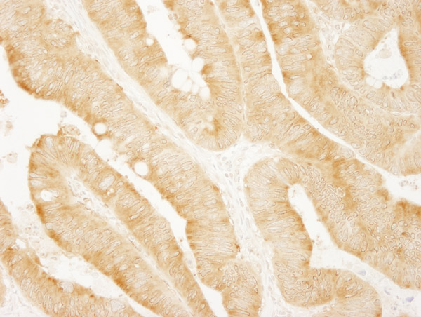 Immunohistochemistry (Formalin/PFA-fixed paraffin-embedded sections) analysis of human colon carcinoma tissue labelling PARP4 with ab86066 at 1/200 (1µg/ml). Detection: DAB.
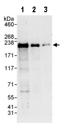 All lanes : Anti-PARP4 antibody (ab86066) at 0.04 µg/mlLane 1 : Hela whole cell lysate at 50 µgLane 2 : Hela whole cell lysate at 15 µgLane 3 : Hela whole cell lysate at 5 µgdeveloped using the ECL technique
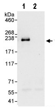 1 mg whole cell lysate from HeLa cells was immunoprecipitated using 3 µg of ab86066. 20% of IP was loaded in each lane and probed with ab86066 at 0.4 µg/ml (lane 1) or with a control IgG (lane 2). Detection: chemiluminescence with an exposure time of 30 seconds.