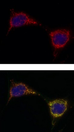
Upper image shows: Immunocytochemistry analysis using ab110314 at 5µg/ml staining PCB in Human fibroblast cells (4% paraformaldehyde fixed and 0.1% Triton X-100 permeabilized) followed by Alexa Fluor® 594 goat anti-mouse IgG (H+L) used at a 1/1000 dilution for 1 hour (red). Note: The target protein locates to the mitochondrial matrix. Lower image shows cells co-stained with an antibody against PDH (green), an enzyme also located in the mitochondrial matrix. The composite image shows an identical mitochondrial pattern for both antibodies indicated by merged orange color.

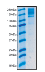
Detection of ab110314 by immunoprecipitation staining of 130kDa PCB in human liver lysate.

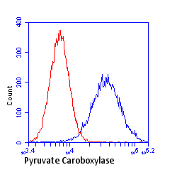
Flow cytometric analysis using ab110314 at 1µg/ml staining PCB in HeLa cells (blue). Isotype control antibody (red).


