PC

Gene Symbol PC
Entrez Gene 338471
Alt Symbol -
Species Bovine
Gene Type protein-coding
Description pyruvate carboxylase
Other Description PCB|pyruvate carboxylase, mitochondrial|pyruvic carboxylase
Swissprots Q866R1 Q29RK2
Accessions DAA13691 Q29RK2 AY185595 AAO27903 AY185596 AAO27904 AY185597 AAO27905 AY185598 AAO27906 AY185599 AAO27907 AY185600 AAO27908 BC114135 AAI14136 XM_005226987 XP_005227044 XM_005226988 XP_005227045 XM_005226989 XP_005227046 XM_005226990 XP_005227047 XM_005226991 XP_005227048 XM_005226992 XP_005227049 XM_005226993 XP_005227050 XM_005226994 XP_005227051 XM_010821049 XP_010819351 XM_010821050 XP_010819352 NM_177946 NP_808815
Function Pyruvate carboxylase catalyzes a 2-step reaction, involving the ATP-dependent carboxylation of the covalently attached biotin in the first step and the transfer of the carboxyl group to pyruvate in the second. Catalyzes in a tissue specific manner, the initial reactions of glucose (liver, kidney) and lipid (adipose tissue, liver, brain) synthesis from pyruvate (By similarity). {ECO:0000250}.
Subcellular Location Mitochondrion matrix {ECO:0000250}.
Top Pathways Citrate cycle (TCA cycle), Pyruvate metabolism, Biosynthesis of amino acids, Carbon metabolism

Anti-PCB antibody - ab77376 from Abcam

Prices $378.00
Sizes 100 µg
Host Goat
Clonality Polyclonal
Anti-PCB antibody (ab77376) at 0.03 µg/ml + Mouse Liver lysate (in RIPA buffer) at 35 µg

Anti-PCB antibody - ab115579 from Abcam

Prices $378.00
Sizes 50 µg
Host Goat
Clonality Polyclonal
ab115579, at 5µg/ml, staining PCB in Formalin-fixed, Paraffin-embedded Human Liver tissue by Immunohistochemistry followed by biotinylated secondary antibody, alkaline phosphatase-streptavidin and chromogen.

Anti-PCB antibody - ab196277 from Abcam

Prices $376.00
Sizes 250 µg
Host Rabbit
Clonality Polyclonal
All lanes : Anti-PCB antibody (ab196277) at 1/500 dilutionLane 1 : Raji cell lysateLane 2 : HEK293 cell lysateLane 3 : 293T cell lysate

Anti-PCB antibody [3H2AD9] - ab110314 from Abcam

Prices $384.00
Sizes 100 µg
Host Mouse
Clonality Monoclonal
Clone 3H2AD9
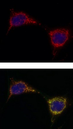
Upper image shows: Immunocytochemistry analysis using ab110314 at 5µg/ml staining PCB in Human fibroblast cells (4% paraformaldehyde fixed and 0.1% Triton X-100 permeabilized) followed by Alexa Fluor® 594 goat anti-mouse IgG (H+L) used at a 1/1000 dilution for 1 hour (red).  Note: The target protein locates to the mitochondrial matrix. Lower image shows cells co-stained with an antibody against PDH (green), an enzyme also located in the mitochondrial matrix. The composite image shows an identical mitochondrial pattern for both antibodies indicated by merged orange color.
Search more