![Anti-PCK2 antibody [EPR14224] - C-terminal (ab187145) at 1/10000 dilution + HepG2 lysate at 20 µgSecondaryGoat Anti-Rabbit IgG, (H+L), Peroxidase conjugated at 1/1000 dilution](http://www.bioprodhub.com/system/product_images/ab_products/2/sub_4/8409_ab187145-220327-ab187145.jpg)
Anti-PCK2 antibody [EPR14224] - C-terminal (ab187145) at 1/10000 dilution + HepG2 lysate at 20 µgSecondaryGoat Anti-Rabbit IgG, (H+L), Peroxidase conjugated at 1/1000 dilution
![All lanes : Anti-PCK2 antibody [EPR14224] - C-terminal (ab187145) at 1/1000 dilutionLane 1 : MCF7 lysateLane 2 : Hela lysateLysates/proteins at 20 µg per lane.SecondaryGoat Anti-Rabbit IgG, (H+L), Peroxidase conjugated at 1/1000 dilution](http://www.bioprodhub.com/system/product_images/ab_products/2/sub_4/8410_ab187145-220325-ab187145b.jpg)
All lanes : Anti-PCK2 antibody [EPR14224] - C-terminal (ab187145) at 1/1000 dilutionLane 1 : MCF7 lysateLane 2 : Hela lysateLysates/proteins at 20 µg per lane.SecondaryGoat Anti-Rabbit IgG, (H+L), Peroxidase conjugated at 1/1000 dilution
![All lanes : Anti-PCK2 antibody [EPR14224] - C-terminal (ab187145) at 1/1000 dilutionLane 1 : Mouse brain lysateLane 2 : Mouse kidney lysateLane 3 : Mouse spleen lysateLane 4 : Rat brain lysateLane 5 : Rat kidney lysateLane 6 : Rat spleen lysateLysates/proteins at 10 µg per lane.SecondaryGoat Anti-Rabbit IgG, (H+L), Peroxidase conjugated at 1/1000 dilution](http://www.bioprodhub.com/system/product_images/ab_products/2/sub_4/8411_ab187145-220322-ab187145c.jpg)
All lanes : Anti-PCK2 antibody [EPR14224] - C-terminal (ab187145) at 1/1000 dilutionLane 1 : Mouse brain lysateLane 2 : Mouse kidney lysateLane 3 : Mouse spleen lysateLane 4 : Rat brain lysateLane 5 : Rat kidney lysateLane 6 : Rat spleen lysateLysates/proteins at 10 µg per lane.SecondaryGoat Anti-Rabbit IgG, (H+L), Peroxidase conjugated at 1/1000 dilution

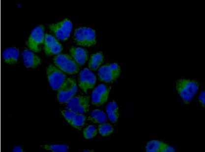
Immunofluorescence analysis of 4% paraformaldehyde-fixed HeLa cells, labeling PCK2 (green) with ab187145 at 1/500 dilution. Alexa Fluor®488-conjugated goat anti-rabbit IgG was used as a secondary antibody at 1/200 dilution. Nuclei were counterstained with DAPI (blue).

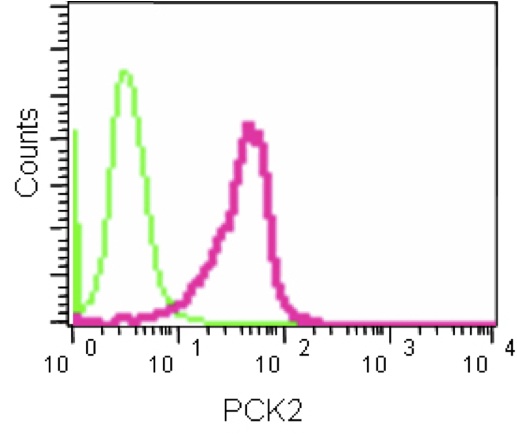
Flow cytometry analysis of PCK2 expression in 2% paraformaldehyde-fixed HeLa cells using ab187145 at 1/50 dilution (red) and a rabbit IgG as negative control (green). A FITC-conjugated goat anti-rabbit IgG (1/150) was used as the secondary antibody.

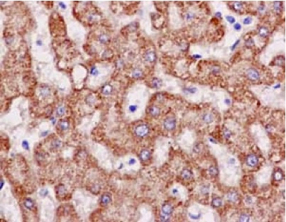
Immunohistochemical analysis of paraffin-embedded Human liver tissue, labeling PCK2 with ab187145 at 1/500 dilution. Detected using HRP Polymer for Rabbit IgG and counter-stained using hematoxylin.
![Anti-PCK2 antibody [EPR14224] - C-terminal (ab187145) at 1/10000 dilution + HepG2 lysate at 20 µgSecondaryGoat Anti-Rabbit IgG, (H+L), Peroxidase conjugated at 1/1000 dilution](http://www.bioprodhub.com/system/product_images/ab_products/2/sub_4/8409_ab187145-220327-ab187145.jpg)
![All lanes : Anti-PCK2 antibody [EPR14224] - C-terminal (ab187145) at 1/1000 dilutionLane 1 : MCF7 lysateLane 2 : Hela lysateLysates/proteins at 20 µg per lane.SecondaryGoat Anti-Rabbit IgG, (H+L), Peroxidase conjugated at 1/1000 dilution](http://www.bioprodhub.com/system/product_images/ab_products/2/sub_4/8410_ab187145-220325-ab187145b.jpg)
![All lanes : Anti-PCK2 antibody [EPR14224] - C-terminal (ab187145) at 1/1000 dilutionLane 1 : Mouse brain lysateLane 2 : Mouse kidney lysateLane 3 : Mouse spleen lysateLane 4 : Rat brain lysateLane 5 : Rat kidney lysateLane 6 : Rat spleen lysateLysates/proteins at 10 µg per lane.SecondaryGoat Anti-Rabbit IgG, (H+L), Peroxidase conjugated at 1/1000 dilution](http://www.bioprodhub.com/system/product_images/ab_products/2/sub_4/8411_ab187145-220322-ab187145c.jpg)


