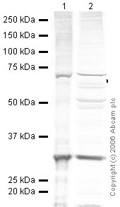
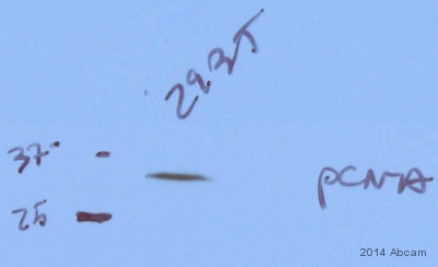
Anti-PCNA antibody
| Name | Anti-PCNA antibody |
|---|---|
| Supplier | Abcam |
| Catalog | ab2426 |
| Prices | $400.00 |
| Sizes | 1 ml |
| Host | Rabbit |
| Clonality | Polyclonal |
| Isotype | IgG |
| Applications | IHC-F IP WB IHC-P ICC/IF ICC/IF |
| Species Reactivities | Mouse, Rat, Hamster, Dog, Human, Zebrafish, Marmoset, Fish |
| Antigen | Synthetic peptide: DMGHLKYYLAPKIEDEEGS , corresponding to C terminal amino acids 243-261 of Human PCNA |
| Description | Rabbit Polyclonal |
| Gene | PCNA |
| Conjugate | Unconjugated |
| Supplier Page | Shop |
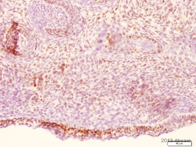
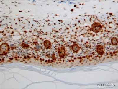
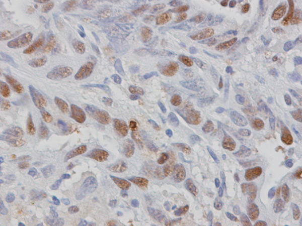
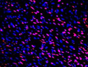
Product images
Product References
IL-15 deficient tax mice reveal a role for IL-1alpha in tumor immunity. - IL-15 deficient tax mice reveal a role for IL-1alpha in tumor immunity.
Rauch DA, Harding JC, Ratner L. PLoS One. 2014 Jan 8;9(1):e85028.
Quantification of healthy and atretic germ cells and follicles in the developing - Quantification of healthy and atretic germ cells and follicles in the developing
Inserra PI, Leopardo NP, Willis MA, Freysselinard AL, Vitullo AD. Reproduction. 2013 Dec 20;147(2):199-209.
Chemopreventive evaluation of a Schiff base derived copper (II) complex against - Chemopreventive evaluation of a Schiff base derived copper (II) complex against
Hajrezaie M, Hassandarvish P, Moghadamtousi SZ, Gwaram NS, Golbabapour S, Najihussien A, Almagrami AA, Zahedifard M, Rouhollahi E, Karimian H, Fani S, Kamalidehghan B, Majid NA, Ali HM, Abdulla MA. PLoS One. 2014 Mar 11;9(3):e91246.
Olive oil and omega-3 polyunsaturated fatty acids suppress intestinal polyp - Olive oil and omega-3 polyunsaturated fatty acids suppress intestinal polyp
Barone M, Notarnicola M, Caruso MG, Scavo MP, Viggiani MT, Tutino V, Polimeno L, Pesetti B, Di Leo A, Francavilla A. Carcinogenesis. 2014 Jul;35(7):1613-9.
Emodin suppresses hyperglycemia-induced proliferation and fibronectin expression - Emodin suppresses hyperglycemia-induced proliferation and fibronectin expression
Gao J, Wang F, Wang W, Su Z, Guo C, Cao S. PLoS One. 2014 Apr 1;9(4):e93588.
Poldip2 knockout results in perinatal lethality, reduced cellular growth and - Poldip2 knockout results in perinatal lethality, reduced cellular growth and
Brown DI, Lassegue B, Lee M, Zafari R, Long JS, Saavedra HI, Griendling KK. PLoS One. 2014 May 5;9(5):e96657.
Role of microRNAs in resveratrol-mediated mitigation of colitis-associated - Role of microRNAs in resveratrol-mediated mitigation of colitis-associated
Altamemi I, Murphy EA, Catroppo JF, Zumbrun EE, Zhang J, McClellan JL, Singh UP, Nagarkatti PS, Nagarkatti M. J Pharmacol Exp Ther. 2014 Jul;350(1):99-109.
Deficiency of the NR4A orphan nuclear receptor NOR1 in hematopoietic stem cells - Deficiency of the NR4A orphan nuclear receptor NOR1 in hematopoietic stem cells
Qing H, Liu Y, Zhao Y, Aono J, Jones KL, Heywood EB, Howatt D, Binkley CM, Daugherty A, Liang Y, Bruemmer D. Stem Cells. 2014 Sep;32(9):2419-29.
R-spondin 2 signalling mediates susceptibility to fatal infectious diarrhoea. - R-spondin 2 signalling mediates susceptibility to fatal infectious diarrhoea.
Papapietro O, Teatero S, Thanabalasuriar A, Yuki KE, Diez E, Zhu L, Kang E, Dhillon S, Muise AM, Durocher Y, Marcinkiewicz MM, Malo D, Gruenheid S. Nat Commun. 2013;4:1898.
An IP3R3- and NPY-expressing microvillous cell mediates tissue homeostasis and - An IP3R3- and NPY-expressing microvillous cell mediates tissue homeostasis and
Jia C, Hayoz S, Hutch CR, Iqbal TR, Pooley AE, Hegg CC. PLoS One. 2013;8(3):e58668.
