![ab193963 staining PCNA in HeLa cells. The cells were fixed with 100% methanol (10 min) and then blocked in 1% BSA/10% normal goat serum/0.3M glycine in 0.1% PBS-Triton X-100 for 1hr. The cells were then incubated with ab193963 at a working dilution of 1/100 (shown in green) and ab7291 (Mouse monoclonal [DM1A] to alpha Tubulin) at 1µg/ml overnight at +4°C, followed by a further incubation at room temperature for 1hr with an AlexaFluor® 594 Goat anti-mouse IgG (H&L - preadsorbed) secondary (ab150120) at 2 μg/ml (shown in pseudo-color red). Nuclear DNA was labelled in blue with DAPI.Image was taken with a Confocal microscope (Leica micro-systems, TCS SP8).](http://www.bioprodhub.com/system/product_images/ab_products/2/sub_4/8527_ab193963-232871-AL488ab1939635ugHeLaMEOHCFM.jpg)
ab193963 staining PCNA in HeLa cells. The cells were fixed with 100% methanol (10 min) and then blocked in 1% BSA/10% normal goat serum/0.3M glycine in 0.1% PBS-Triton X-100 for 1hr. The cells were then incubated with ab193963 at a working dilution of 1/100 (shown in green) and ab7291 (Mouse monoclonal [DM1A] to alpha Tubulin) at 1µg/ml overnight at +4°C, followed by a further incubation at room temperature for 1hr with an AlexaFluor® 594 Goat anti-mouse IgG (H&L - preadsorbed) secondary (ab150120) at 2 μg/ml (shown in pseudo-color red). Nuclear DNA was labelled in blue with DAPI.Image was taken with a Confocal microscope (Leica micro-systems, TCS SP8).
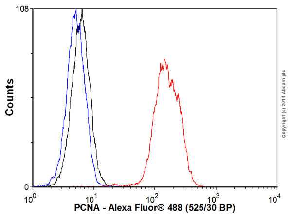
Overlay histogram showing HeLa cells stained with ab193963 (red line). The cells were fixed with 80% methanol (5 min) and then permeabilized with 0.1% PBS-Tween for 20 min. The cells were then incubated in 1x PBS / 10% normal goat serum / 0.3M glycine to block non-specific protein-protein interactions followed by the antibody (ab193963, 0.1μg/1x106 cells) for 30 min at 22°C. Isotype control antibody (black line) was rabbit IgG (monoclonal) (Alexa Fluor® 488) (0.2μl/1x106 cells) for 30 min at 22ºC. Unlabelled sample (blue line) was also used as a control.Acquisition of >5,000 events were collected using a 20mW Argon ion laser (488nm) and 525/30 bandpass filter.