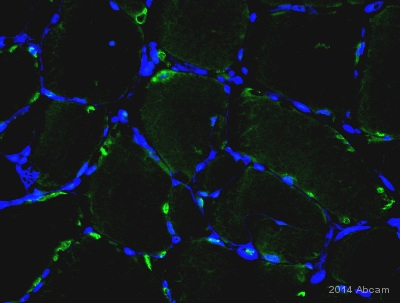
ab23914 staining PDGF BB (green) in mouse skeletal muscle tissue sections by Immunohistochemistry (IHC-P - paraformaldehyde-fixed, paraffin-embedded sections). Tissue was fixed with formaldehyde and blocked with 10% serum for 10 minutes at 25°C; antigen retrieval was by heat mediation in Tris pH 9. Samples were incubated with primary antibody (1/500 in antibody diluent reagent) for 14 hours at 4°C. An Alexa Fluor® 647-conjugated goat anti-rabbit IgG polyclonal (1/200) was used as the secondary antibody. DAPI (blue) - nuclei.See Abreview
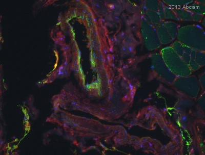
ab23914 staining PDGF BB (green) in Mouse skeletal muscle (gastrocnemius) tissue sections by Immunohistochemistry (IHC-P - paraformaldehyde-fixed, paraffin-embedded sections). Tissue was fixed with methacarn and blocked with 10% serum for 10 minutes at 25°C; antigen retrieval was by heat mediation in Tris pH 8. Samples were incubated with primary antibody (1/250 in antibody diluent reagent) for 14 hours at 4°C. An Alexa Fluor® 488-conjugated Goat anti-rabbit IgG polyclonal (1/200) was used as the secondary antibody. Red - WGA membranes, Blue - DAPI Nuclei.See Abreview
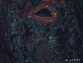
ab23914 staining PDGF BB in Human skeletal muscle (gastrocnemius) tissue sections by Immunohistochemistry (IHC-Fr - frozen sections). Tissue was fixed with formaldehyde and blocked with 10% serum for 20 minutes at 25°C. Samples were incubated with primary antibody (1/150) for 14 hours at 4°C. An Alexa Fluor® 555-conjugated Goat anti-rabbit IgG polyclonal (1/5000) was used as the secondary antibody.See Abreview

All lanes : Anti-PDGF BB antibody (ab23914) at 1 µg/mlLane 1 : Active PDGF BB full length protein (ab9706)Lane 2 : Active PDGF BB full length protein (ab9706) with Human PDGF BB peptide (ab24035) at 1 µgLysates/proteins at 0.01 µg per lane.SecondaryGoat polyclonal to Rabbit IgG (Alexa Fluor® 680) at 1/10000 dilutionPerformed under reducing conditions.

Image courtesy of Human Protein Atlasab23914 staining PDGF BB in Human kidney (male). The paraffin embedded tissue was incubated with ab23914 (1/500 dilution) for 30 mins at room temperature. Antigen retrieval was performed by heat induction in citrate buffer pH 6. ab23914 was tested in a tissue microarray (TMA) containing a wide range of normal and cancer tissues as well as a cell microarray con
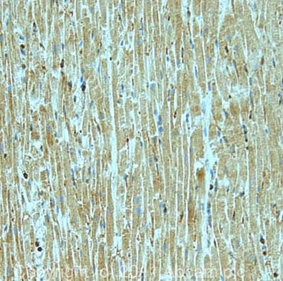
IHC image of PDGF BB staining in human heart formalin fixed paraffin embedded tissue section, performed on a Leica BondTM system using the standard protocol F. The section was pre-treated using heat mediated antigen retrieval with sodium citrate buffer (pH6, epitope retrieval solution 1) for 20 mins. The section was then incubated with ab23914, 5µg/ml, for 15 mins at room temperature and detected using an HRP conjugated compact polymer system. DAB was used as the chromogen. The section was then counterstained with haematoxylin and mounted with DPX.
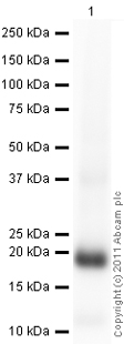
developed using the ECL techniquePerformed under reducing conditions.
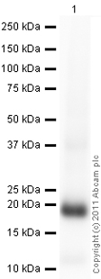
developed using the ECL techniquePerformed under reducing conditions.
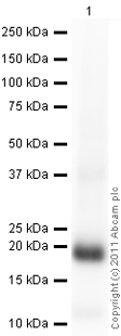
developed using the ECL techniquePerformed under reducing conditions.


