Anti-PDK4 antibody
| Name | Anti-PDK4 antibody |
|---|---|
| Supplier | Abcam |
| Catalog | ab38242 |
| Host | Rabbit |
| Clonality | Polyclonal |
| Isotype | IgG |
| Applications | IHC-P ICC/IF ICC/IF ELISA WB |
| Species Reactivities | Mouse, Human |
| Antigen | Synthetic peptide corresponding to Human PDK4 aa 382-410 (C terminal) conjugated to Keyhole Limpet Haemocyanin (KLH) |
| Description | Rabbit Polyclonal |
| Gene | PDK4 |
| Conjugate | Unconjugated |
| Supplier Page | Shop |
Product images
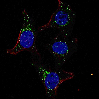 Fluorescent confocal image of HeLa cells stained with PDK4 (ab38242) antibody. HeLa cells were fixed with 4% PFA (20 min), permeabilized with Triton X-100 (0.2%, 30 min). Cells were then incubated with ab38242 PDK4 () primary antibody (1:100, 2 h at room temperature). For secondary antibody, Alexa Fluor® 488 conjugated donkey anti-rabbit antibody (green) was used (1:1000, 1h). Nuclei were counterstained with Hoechst 33342 (blue) (10 μg/ml, 5 min). Note the highly specific localization of the PDK4 mainly to the cytoplasm.
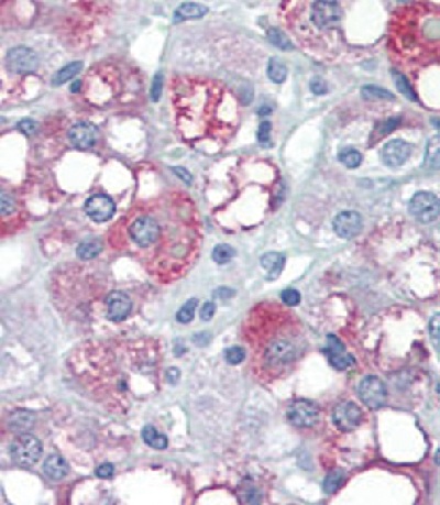 Immunohistochemical staining of PDK4 in Human adrenal tissue sections (IHC-P - paraformaldehyde-fixed, paraffin-embedded sections) with ab38242 at a dilution of 1/25. Tissue was fixed with formaldehyde and blocked with 3% BSA for 0.5 hour at 38°C. Antigen retrieval was heat mediation with a citrate buffer (pH6). Samples were incubated with primary antibody (1/25) for 1 hours at 37°C. A HRP-conjugated goat anti-rabbit polyclonal (ready to use) was used as the secondary antibody.
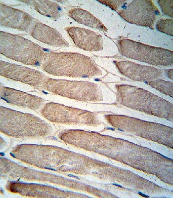 Immunohistochemical staining of PDK4 in Human skeletal muscle sections (IHC-P - paraformaldehyde-fixed, paraffin-embedded sections) with ab38242. Tissue was fixed with formaldehyde and blocked with 3% BSA for 0.5 hour at 38°C. Antigen retrieval was heat mediation with a citrate buffer (pH6). Samples were incubated with primary antibody (1/25) for 1 hours at 37°C. A HRP-conjugated goat anti-rabbit polyclonal (ready to use) was used as the secondary antibody.
 Anti-PDK4 antibody (ab38242) at 1/1000 dilution + mouse skeletal muscle tissue lysateSecondaryHRP goat anti-rabbit (H+L) at 1/5000 dilution
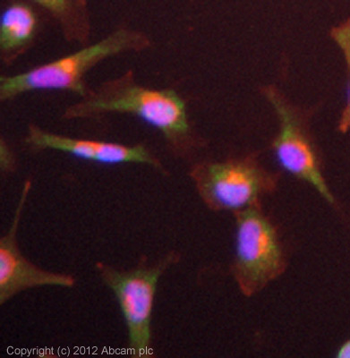 ICC/IF image of ab38242 stained HeLa cells. The cells were 4% formaldehyde (10 min) and then incubated in 1%BSA / 10% normal goat serum / 0.3M glycine in 0.1% PBS-Tween for 1h to permeabilise the cells and block non-specific protein-protein interactions. The cells were then incubated with the antibody (ab38242, 10µg/ml) overnight at +4°C. The secondary antibody (green) was for 1h. Alexa Fluor® 594 WGA was used to label plasma membranes (red) at a 1/200 dilution for 1h. DAPI was used to stain the cell nuclei (blue) at a concentration of 1.43µM.
Product References
Proteome-based systems biology analysis of the diabetic mouse aorta reveals major - Proteome-based systems biology analysis of the diabetic mouse aorta reveals major
Husi H, Van Agtmael T, Mullen W, Bahlmann FH, Schanstra JP, Vlahou A, Delles C, Perco P, Mischak H. Circ Cardiovasc Genet. 2014 Apr;7(2):161-70. doi:
FAT/CD36 regulates PEPCK expression in adipose tissue. - FAT/CD36 regulates PEPCK expression in adipose tissue.
Wan Z, Matravadia S, Holloway GP, Wright DC. Am J Physiol Cell Physiol. 2013 Mar 1;304(5):C478-84. doi:
IL-6 indirectly modulates the induction of glyceroneogenic enzymes in adipose - IL-6 indirectly modulates the induction of glyceroneogenic enzymes in adipose
Wan Z, Ritchie I, Beaudoin MS, Castellani L, Chan CB, Wright DC. PLoS One. 2012;7(7):e41719.
Impaired cardiac functional reserve in type 2 diabetic db/db mice is associated - Impaired cardiac functional reserve in type 2 diabetic db/db mice is associated
Daniels A, van Bilsen M, Janssen BJ, Brouns AE, Cleutjens JP, Roemen TH, Schaart G, van der Velden J, van der Vusse GJ, van Nieuwenhoven FA. Acta Physiol (Oxf). 2010 Sep;200(1):11-22.
miRNA in the regulation of skeletal muscle adaptation to acute endurance exercise - miRNA in the regulation of skeletal muscle adaptation to acute endurance exercise
Safdar A, Abadi A, Akhtar M, Hettinga BP, Tarnopolsky MA. PLoS One. 2009;4(5):e5610.