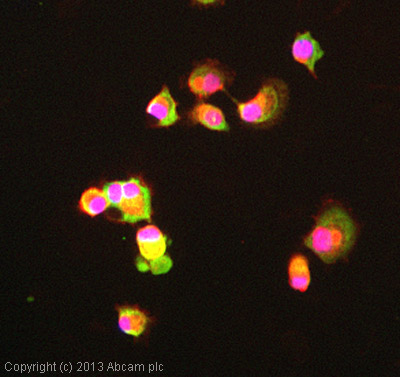
ICC/IF image of ab98298 stained Panc-1 cells. The cells were 4% formaldehyde fixed (10 min) and then incubated in 1%BSA / 10% normal goat serum / 0.3M glycine in 0.1% PBS-Tween for 1h to permeabilise the cells and block non-specific protein-protein interactions. The cells were then incubated with the antibody (ab98298, 5µg/ml) overnight at +4°C. The secondary antibody (green) was ab96899, DyLight® 488 goat anti-rabbit IgG (H+L) used at a 1/250 dilution for 1h. Alexa Fluor® 594 WGA was used to label plasma membranes (red) at a 1/200 dilution for 1h. DAPI was used to stain the cell nuclei (blue) at a concentration of 1.43µM
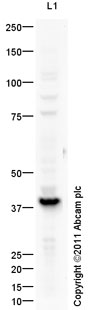
Anti-PDX1 antibody (ab98298) at 1 µg/ml + Human Pancreatic Islet Cells at 10 µgSecondaryGoat Anti-Rabbit IgG H&L (HRP) preadsorbed (ab97080) at 1/5000 dilutiondeveloped using the ECL techniquePerformed under reducing conditions.
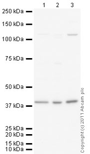
All lanes : Anti-PDX1 antibody (ab98298) at 1 µg/mlLane 1 : HeLa (Human epithelial carcinoma cell line) Nuclear Lysate Lane 2 : Jurkat (Human T cell lymphoblast-like cell line) Nuclear Lysate (ab14844)Lane 3 : Pancreas (Mouse) Tissue Lysate Lysates/proteins at 10 µg per lane.SecondaryGoat Anti-Rabbit IgG H&L (HRP) preadsorbed (ab97080) at 1/5000 dilutiondeveloped using the ECL techniquePerformed under reducing conditions.
![PDX1 was immunoprecipitated using 0.5mg Hela whole cell extract, 5µg of Rabbit polyclonal to PDX1 and 50µl of protein G magnetic beads (+). No antibody was added to the control (-). The antibody was incubated under agitation with Protein G beads for 10min, Hela whole cell extract lysate diluted in RIPA buffer was added to each sample and incubated for a further 10min under agitation.Proteins were eluted by addition of 40µl SDS loading buffer and incubated for 10min at 70oC; 10µl of each sample was separated on a SDS PAGE gel, transferred to a nitrocellulose membrane, blocked with 5% BSA and probed with ab98298.Secondary: Mouse monoclonal [SB62a] Secondary Antibody to Rabbit IgG light chain (HRP) (ab99697).Band: 36kDa: PDX1.](http://www.bioprodhub.com/system/product_images/ab_products/2/sub_4/9519_PDX1-Primary-antibodies-ab98298-5.jpg)
PDX1 was immunoprecipitated using 0.5mg Hela whole cell extract, 5µg of Rabbit polyclonal to PDX1 and 50µl of protein G magnetic beads (+). No antibody was added to the control (-). The antibody was incubated under agitation with Protein G beads for 10min, Hela whole cell extract lysate diluted in RIPA buffer was added to each sample and incubated for a further 10min under agitation.Proteins were eluted by addition of 40µl SDS loading buffer and incubated for 10min at 70oC; 10µl of each sample was separated on a SDS PAGE gel, transferred to a nitrocellulose membrane, blocked with 5% BSA and probed with ab98298.Secondary: Mouse monoclonal [SB62a] Secondary Antibody to Rabbit IgG light chain (HRP) (ab99697).Band: 36kDa: PDX1.