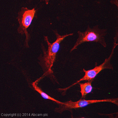
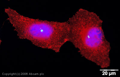
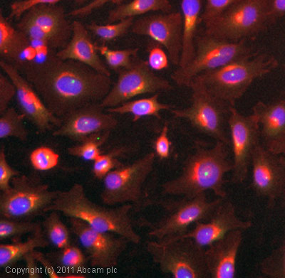
Anti-Pericentrin antibody - Centrosome Marker
| Name | Anti-Pericentrin antibody - Centrosome Marker |
|---|---|
| Supplier | Abcam |
| Catalog | ab4448 |
| Prices | $394.00 |
| Sizes | 100 µg |
| Host | Rabbit |
| Clonality | Polyclonal |
| Isotype | IgG |
| Applications | ICC/IF ICC/IF IHC-P ICC/IF IHC-F |
| Species Reactivities | Mouse, Rat, Rabbit, Human, Monkey |
| Antigen | The pericentrin clone used is 1 |
| Description | Rabbit Polyclonal |
| Gene | PCNT |
| Conjugate | Unconjugated |
| Supplier Page | Shop |
Product images
Product References
TACC3-ch-TOG track the growing tips of microtubules independently of clathrin and - TACC3-ch-TOG track the growing tips of microtubules independently of clathrin and
Gutierrez-Caballero C, Burgess SG, Bayliss R, Royle SJ. Biol Open. 2015 Jan 16;4(2):170-9.
Interactions between lens epithelial and fiber cells reveal an intrinsic - Interactions between lens epithelial and fiber cells reveal an intrinsic
Dawes LJ, Sugiyama Y, Lovicu FJ, Harris CG, Shelley EJ, McAvoy JW. Dev Biol. 2014 Jan 15;385(2):291-303.
C2orf62 and TTC17 are involved in actin organization and ciliogenesis in - C2orf62 and TTC17 are involved in actin organization and ciliogenesis in
Bontems F, Fish RJ, Borlat I, Lembo F, Chocu S, Chalmel F, Borg JP, Pineau C, Neerman-Arbez M, Bairoch A, Lane L. PLoS One. 2014 Jan 27;9(1):e86476.
CERKL, a retinal disease gene, encodes an mRNA-binding protein that localizes in - CERKL, a retinal disease gene, encodes an mRNA-binding protein that localizes in
Fathinajafabadi A, Perez-Jimenez E, Riera M, Knecht E, Gonzalez-Duarte R. PLoS One. 2014 Feb 3;9(2):e87898.
C2cd3 is critical for centriolar distal appendage assembly and ciliary vesicle - C2cd3 is critical for centriolar distal appendage assembly and ciliary vesicle
Ye X, Zeng H, Ning G, Reiter JF, Liu A. Proc Natl Acad Sci U S A. 2014 Feb 11;111(6):2164-9. doi:
Multimodal effects of small molecule ROCK and LIMK inhibitors on mitosis, and - Multimodal effects of small molecule ROCK and LIMK inhibitors on mitosis, and
Oku Y, Tareyanagi C, Takaya S, Osaka S, Ujiie H, Yoshida K, Nishiya N, Uehara Y. PLoS One. 2014 Mar 18;9(3):e92402.
SUN proteins facilitate the removal of membranes from chromatin during nuclear - SUN proteins facilitate the removal of membranes from chromatin during nuclear
Turgay Y, Champion L, Balazs C, Held M, Toso A, Gerlich DW, Meraldi P, Kutay U. J Cell Biol. 2014 Mar 31;204(7):1099-109.
Dido3-dependent HDAC6 targeting controls cilium size. - Dido3-dependent HDAC6 targeting controls cilium size.
Sanchez de Diego A, Alonso Guerrero A, Martinez-A C, van Wely KH. Nat Commun. 2014 Mar 25;5:3500.
STK31 is a cell-cycle regulated protein that contributes to the tumorigenicity of - STK31 is a cell-cycle regulated protein that contributes to the tumorigenicity of
Kuo PL, Huang YL, Hsieh CC, Lee JC, Lin BW, Hung LY. PLoS One. 2014 Mar 25;9(3):e93303.
KDM4C (GASC1) lysine demethylase is associated with mitotic chromatin and - KDM4C (GASC1) lysine demethylase is associated with mitotic chromatin and
Kupershmit I, Khoury-Haddad H, Awwad SW, Guttmann-Raviv N, Ayoub N. Nucleic Acids Res. 2014 Jun;42(10):6168-82.


