Anti-PHD3 antibody
| Name | Anti-PHD3 antibody |
|---|---|
| Supplier | Abcam |
| Catalog | ab30782 |
| Prices | $398.00 |
| Sizes | 100 µg |
| Host | Rabbit |
| Clonality | Polyclonal |
| Isotype | IgG |
| Applications | IHC-P IP ICC/IF ICC/IF WB |
| Species Reactivities | Mouse, Rat, Human |
| Antigen | Synthetic peptide conjugated to KLH derived from within residues 50 - 150 of Human PHD3/prolyl hydroxylase |
| Description | Rabbit Polyclonal |
| Gene | EGLN3 |
| Conjugate | Unconjugated |
| Supplier Page | Shop |
Product images
 All lanes : Anti-PHD3 antibody (ab30782) at 1 µg/mlLane 1 : Hela-Vehicle treated (Negative Control) Whole Cell Lysate (ab116321)Lane 2 : Hela-DFO treated (0.5mM, 24h) Whole Cell Lysate (ab116322)Lane 3 : Human HeLa Nuclear DFO treatedLane 4 : Human HeLa Cytoplasmic DFO treatedLysates/proteins at 20 µg per lane.SecondaryGoat Anti-Rabbit IgG H&L (HRP) (ab97051) at 1/10000 dilutiondeveloped using the ECL techniquePerformed under reducing conditions.
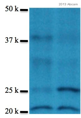 All lanes : Anti-PHD3 antibody (ab30782) at 1/1000 dilutionLane 1 : Mouse embroyonic fibroblast cell lysate. Treated 21% O2 for 24 hoursLane 2 : Mouse embroyonic fibroblast cell lysate. Treated 1% O2 for 24 hoursLysates/proteins at 5 µg per lane.SecondaryGoat polyclonal anti-rabbit HRP-conjugate at 1/10000 dilutiondeveloped using the ECL techniquePerformed under reducing conditions.
 ab30782 immunoprecipitating PHD3 from rat PC12 (whole cell lysate - the cells were either normoxia (21% oxygen) or hypoxia (1% hypoxia). A protein A matrix was used with the antibody at a dilution of 1/1000. A non-Abcam antibody was used in the subsequent western blot. The doublet observed is specific to PHD3 - most probably a modified form or an alternately spliced form. Note: This antibody IP's a hypoxia inducible species of ~27 kDa and ~30 kDa. This is the same molecular weight as the human and mouse isoform, not the SM-20 variant with the N-terminal mitochondrial leader sequence. Lane 1: normoxia - inputLane 2: hypoxia - inputLane 3: normoxia - control antibody IPLane 4: hypoxia - control antibody IPLane 5: normoxia - ab30782 IPLane 6: hypoxia - ab30782 IPSee Abreview
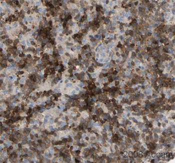 Image courtesy of Human Protein Atlasab30782 staining PHD3 in human lymph node, showing strong staining in the follicle cells of the paracortex. Paraffin embedded human lymph node tissue was incubated with ab30782 (1/300 dilution) for 30 mins at room temperature. Antigen retrieval was performed by heat induction in citrate buffer pH 6.ab30782 was tested in a tissue microarray (TMA) containing a wide range of normal and cancer tissues as well as a cell microarray consisting of a range of commonly used, well characterised human cell lines. Further results for this antibody can be found at www.proteinatlas.org.
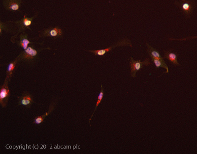 ICC/IF image of ab30782 stained HepG2 cells. The cells were 4% formaldehyde fixed (10 min) and then incubated in 1%BSA / 10% normal goat serum / 0.3M glycine in 0.1% PBS-Tween for 1h to permeabilise the cells and block non-specific protein-protein interactions. The cells were then incubated with the antibody ab30782 at 5µg/ml overnight at +4°C. The secondary antibody (green) was DyLight® 488 goat anti- rabbit (ab96899) IgG (H+L) used at a 1/1000 dilution for 1h. Alexa Fluor® 594 WGA was used to label plasma membranes (red) at a 1/200 dilution for 1h. DAPI was used to stain the cell nuclei (blue) at a concentration of 1.43µM.
Product References
Differential expression of prolyl hydroxylase 1 in patients with ulcerative - Differential expression of prolyl hydroxylase 1 in patients with ulcerative
Van Welden S, Laukens D, Ferdinande L, De Vos M, Hindryckx P. J Inflamm (Lond). 2013 Nov 20;10(1):36.
The prolyl hydroxylase PHD3 identifies proinflammatory macrophages and its - The prolyl hydroxylase PHD3 identifies proinflammatory macrophages and its
Escribese MM, Sierra-Filardi E, Nieto C, Samaniego R, Sanchez-Torres C, Matsuyama T, Calderon-Gomez E, Vega MA, Salas A, Sanchez-Mateos P, Corbi AL. J Immunol. 2012 Aug 15;189(4):1946-54.
Se-methylselenocysteine sensitizes hypoxic tumor cells to irinotecan by targeting - Se-methylselenocysteine sensitizes hypoxic tumor cells to irinotecan by targeting
Chintala S, Toth K, Cao S, Durrani FA, Vaughan MM, Jensen RL, Rustum YM. Cancer Chemother Pharmacol. 2010 Oct;66(5):899-911. doi:
HIF prolyl hydroxylase-3 mediates alpha-ketoglutarate-induced apoptosis and tumor - HIF prolyl hydroxylase-3 mediates alpha-ketoglutarate-induced apoptosis and tumor
Tennant DA, Gottlieb E. J Mol Med (Berl). 2010 Aug;88(8):839-49.
Activation of negative regulators of the hypoxia-inducible factor (HIF) pathway - Activation of negative regulators of the hypoxia-inducible factor (HIF) pathway
Zolk O, Solbach TF, Eschenhagen T, Weidemann A, Fromm MF. Biochem Biophys Res Commun. 2008 Nov 14;376(2):315-20. doi: