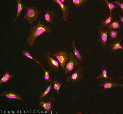
ab109119 stained HeLa cells. The cells were 4% formaldhyde fixed for 10 minutes at room temperature and then incubated in 1%BSA / 10% normal goat serum / 0.3M glycine in 0.1% PBS-Tween for 1hour at room temperature to permeabilise the cells and block non-specific protein-protein interactions. The cells were then incubated with the antibody (ab109119 at 1/100) overnight at +4°C. The secondary antibody (pseudo-colored green) was Goat Anti-Rabbit IgG H&L (Alexa Fluor® 488) preadsorbed (ab150081) used at a 1/1000 dilution for 1hour at room temperature. Alexa Fluor® 594 WGA was used to label plasma membranes (pseudo-colored red) at a 1/200 dilution for 1hour at room temperature. DAPI was used to stain the cell nuclei (pseudo-colored blue) at a concentration of 1.43µM for 1hour at room temperature.
![All lanes : Anti-PIST antibody [EPR4079] (ab109119) at 1/1000 dilutionLane 1 : A375 cell lysateLane 2 : 293T cell lysateLane 3 : Fetal kidney lysateLane 4 : PC-3 cell lysateLane 5 : Human prostate lysateLysates/proteins at 10 µg per lane.](http://www.bioprodhub.com/system/product_images/ab_products/2/sub_4/12225_PIST-Primary-antibodies-ab109119-1.jpg)
All lanes : Anti-PIST antibody [EPR4079] (ab109119) at 1/1000 dilutionLane 1 : A375 cell lysateLane 2 : 293T cell lysateLane 3 : Fetal kidney lysateLane 4 : PC-3 cell lysateLane 5 : Human prostate lysateLysates/proteins at 10 µg per lane.
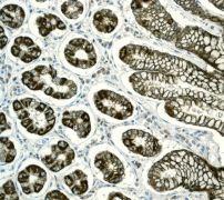
Immunohistochemical staining of PIST expression in paraffin-embedded Human stomach tissue using ab109119 at 1/250 dilution.
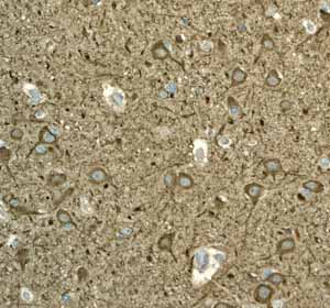
ab109119 showing positive staining in Normal brain tissue.
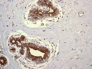
ab109119 showing positive staining in Normal breast tissue.
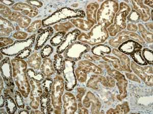
ab109119 showing positive staining in Normal kidney tissue.
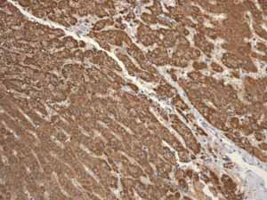
ab109119 showing positive staining in Normal liver tissue.
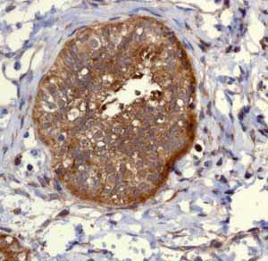
ab109119 showing positive staining in Normal ovary tissue.

![All lanes : Anti-PIST antibody [EPR4079] (ab109119) at 1/1000 dilutionLane 1 : A375 cell lysateLane 2 : 293T cell lysateLane 3 : Fetal kidney lysateLane 4 : PC-3 cell lysateLane 5 : Human prostate lysateLysates/proteins at 10 µg per lane.](http://www.bioprodhub.com/system/product_images/ab_products/2/sub_4/12225_PIST-Primary-antibodies-ab109119-1.jpg)


