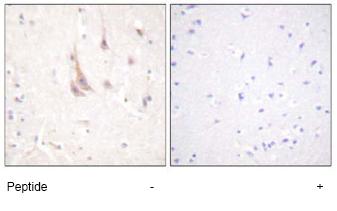
Anti-PKC zeta antibody
| Name | Anti-PKC zeta antibody |
|---|---|
| Supplier | Abcam |
| Catalog | ab59364 |
| Prices | $398.00 |
| Sizes | 100 µg |
| Host | Rabbit |
| Clonality | Polyclonal |
| Isotype | IgG |
| Applications | WB ELISA IHC-P IP ICC/IF ICC/IF |
| Species Reactivities | Mouse, Rat, Human, Xenopus |
| Antigen | Synthesized non-phosphopeptide derived from human PKC zeta around the phosphorylation site of threonine 560 (Q-L-TP-P-D) |
| Description | Rabbit Polyclonal |
| Gene | PRKCZ |
| Conjugate | Unconjugated |
| Supplier Page | Shop |
Product images
Product References
Protein expression of PKCZ (Protein Kinase C Zeta), Munc18c, and Syntaxin-4 in - Protein expression of PKCZ (Protein Kinase C Zeta), Munc18c, and Syntaxin-4 in
Rivero R, Garin CA, Ormazabal P, Silva A, Carvajal R, Gabler F, Romero C, Vega M. Reprod Biol Endocrinol. 2012 Mar 5;10:17.
Cellular mechanism of insulin resistance in nonalcoholic fatty liver disease. - Cellular mechanism of insulin resistance in nonalcoholic fatty liver disease.
Kumashiro N, Erion DM, Zhang D, Kahn M, Beddow SA, Chu X, Still CD, Gerhard GS, Han X, Dziura J, Petersen KF, Samuel VT, Shulman GI. Proc Natl Acad Sci U S A. 2011 Sep 27;108(39):16381-5. doi:
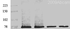
![PKC zeta was immunoprecipitated using 0.5mg Mouse Brain tissue, 5µg of Rabbit polyclonal to PKC zeta and 50µl of protein G magnetic beads (+). No antibody was added to the control (-).The antibody was incubated under agitation with Protein G beads for 10min, Mouse Brain tissue lysate diluted in RIPA buffer was added to each sample and incubated for a further 10min under agitation.Proteins were eluted by addition of 40µl SDS loading buffer and incubated for 10min at 70°C; 10µl of each sample was separated on a SDS PAGE gel, transferred to a nitrocellulose membrane, blocked with 5% BSA and probed with ab59364.Secondary: Mouse monoclonal [SB62a] Secondary Antibody to Anti-Rabbit IgG light chain HRP (ab99697).Band: 68kDa; PKC zeta, non specific bands - 52kDa: We are unsure as to the identity of this extra band.](http://www.bioprodhub.com/system/product_images/ab_products/2/sub_4/12742_ab59364-197839-IPV023ab5936416min.jpg)

