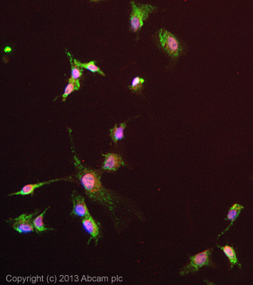
ICC/IF image of ab126203 stained HepG2 cells. The cells were 4% Formaldehyde fixed (10 min) and then incubated in 1%BSA / 10% normal Goat serum / 0.3M glycine in 0.1% PBS-Tween for 1h to permeabilise the cells and block non-specific protein-protein interactions. The cells were then incubated with the antibody (ab126203, 1µg/ml) overnight at +4°C. The secondary antibody (green) was ab96899, DyLight® 488 Goat anti-Rabbit IgG (H+L) used at a 1/250 dilution for 1h. Alexa Fluor® 594 WGA was used to label plasma membranes (red) at a 1/200 dilution for 1h. DAPI was used to stain the cell nuclei (blue) at a concentration of 1.43µM.
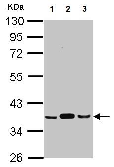
All lanes : Anti-Pyruvate Dehydrogenase E1 beta subunit antibody (ab126203) at 1/1000 dilutionLane 1 : A549 whole cell lysateLane 2 : H1299 whole cell lysateLane 3 : HCT116 whole cell lysateLysates/proteins at 30 µg per lane.
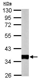
Anti-Pyruvate Dehydrogenase E1 beta subunit antibody (ab126203) at 1/10000 dilution + Mouse heart whole cell lysate at 50 µg
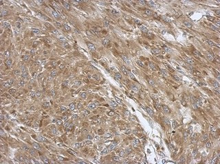
ab126203, at 1/500 dilution, staining Pyruvate Dehydrogenase E1 beta subunit in paraffin-embedded U87 xenograft tissue by Immunohistochemistry.