CDK9 (C12F7) Rabbit mAb
| Name | CDK9 (C12F7) Rabbit mAb |
|---|---|
| Supplier | Cell Signaling Technology |
| Catalog | 2316 |
| Prices | $99.00, $246.00 |
| Sizes | 20 µl (2 western blots), 100 µl (10 western blots) |
| Host | Rabbit |
| Clonality | Monoclonal |
| Isotype | IgG |
| Clone | C12F7 |
| Applications | WB IP IHC-P IHC-F ICC/IF FC |
| Species Reactivities | Human, Mouse, Rat, Hamster, Monkey, Bovine, Dog |
| Antigen | Monoclonal antibody is produced by immunizing animals with a synthetic peptide corresponding to residues near the carboxy terminus of human CDK9. |
| Description | Rabbit Monoclonal |
| Gene | CDK9 |
| Supplier Page | Shop |
Product images
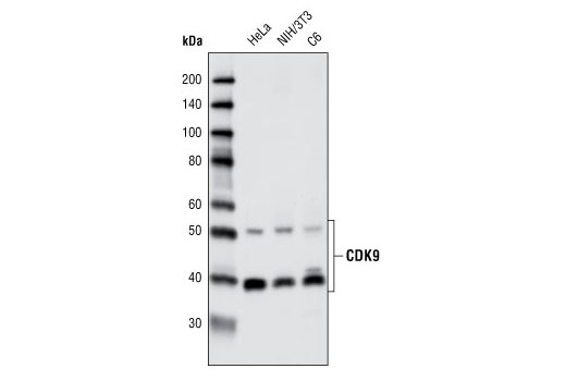 Western blot analysis of extracts from various cell types using CDK9 (C12F7) Rabbit mAb.
 Immunoprecipitation of CDK9 from HeLa cells using CDK9 (C12F7) Rabbit mAb. Western blot detection was performed using the same antibody. Lane 1 is 5% input.
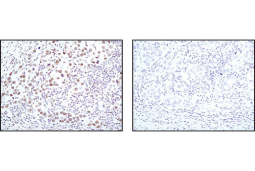 Immunohistochemical analysis of paraffin-embedded human breast carcinoma using CDK9 (C12F7) Rabbit mAb in the presence of control peptide (left) or antigen specific peptide (right).
 Immunohistochemical analysis of paraffin-embedded K7M2 mouse syngeneic tumor using CDK9 (C12F7) Rabbit mAb.
 Immunohistochemical analysis of frozen SKOV-3 xenograft using CDK9 (C12F7) Rabbit mAb.
 Confocal immunofluorescent analysis of HeLa cells using CDK9 (C12F7) Rabbit mAb (green). Actin filaments have been labeled with DY-555 phalloidin (red).
 Flow cytometric analysis of Jurkat cells using CDK9 (C12F7) Rabbit mAb (blue) compared to a nonspecific negative control antibody (red).