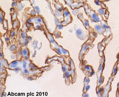
Anti-RAGE antibody
| Name | Anti-RAGE antibody |
|---|---|
| Supplier | Abcam |
| Catalog | ab54741 |
| Prices | $394.00 |
| Sizes | 100 µg |
| Host | Mouse |
| Clonality | Monoclonal |
| Isotype | IgG2a |
| Applications | WB FC IHC-P |
| Species Reactivities | Human |
| Antigen | Recombinant full length protein, corresponding to amino acids 23-405 of Human RAGE |
| Description | Mouse Monoclonal |
| Gene | AGER |
| Conjugate | Unconjugated |
| Supplier Page | Shop |
Product images
Product References
MK615 decreases RAGE expression and inhibits TAGE-induced proliferation in - MK615 decreases RAGE expression and inhibits TAGE-induced proliferation in
Sakuraoka Y, Sawada T, Okada T, Shiraki T, Miura Y, Hiraishi K, Ohsawa T, Adachi M, Takino J, Takeuchi M, Kubota K. World J Gastroenterol. 2010 Nov 14;16(42):5334-41.
Irreversibly glycated LDL induce oxidative and inflammatory state in human - Irreversibly glycated LDL induce oxidative and inflammatory state in human
Toma L, Stancu CS, Botez GM, Sima AV, Simionescu M. Biochem Biophys Res Commun. 2009 Dec 18;390(3):877-82. doi:


