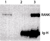
Detection of RANK in RAW cells. Lane 1: mouse cell line. Lane 2 & 3: IP/Western blot analysis of RANK. RANK protein from RAW cell lysate was immunoprecipitated either with control antibody (lane 2) or ab13918 (lane 3), and detected with ab13918.

IHC image of ab13918 staining in human t cell lymphoma formalin fixed paraffin embedded tissue section, performed on a Leica BondTM system using the standard protocol F. The section was pre-treated using heat mediated antigen retrieval with sodium citrate buffer (pH6, epitope retrieval solution 1) for 20 mins. The section was then incubated with ab13918, 5µg/ml, for 15 mins at room temperature and detected using an HRP conjugated compact polymer system. DAB was used as the chromogen. The section was then counterstained with haematoxylin and mounted with DPX. For other IHC staining systems (automated and non-automated) customers should optimize variable parameters such as antigen retrieval conditions, primary antibody concentration and antibody incubation times.