Anti-RANK antibody [9A725]
| Name | Anti-RANK antibody [9A725] |
|---|---|
| Supplier | Abcam |
| Catalog | ab12008 |
| Prices | $390.00 |
| Sizes | 50 µg |
| Host | Mouse |
| Clonality | Monoclonal |
| Isotype | IgG1 |
| Clone | 9A725 |
| Applications | WB ICC/IF ICC/IF FC IHC-P |
| Species Reactivities | Human |
| Antigen | Fusion protein, corresponding to amino acids 326-616 of Human RANK |
| Description | Mouse Monoclonal |
| Gene | TNFRSF11A |
| Conjugate | Unconjugated |
| Supplier Page | Shop |
Product images
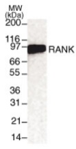 RANK detection by Western blot. The analysis of RANK in 293 cells transfected with RANK cDNA. A single protein band of approximate mol. wt. of 97 kDa was detected by using anti-RANK at 2 ug/ml.
Product References
Vps35 loss promotes hyperresorptive osteoclastogenesis and osteoporosis via - Vps35 loss promotes hyperresorptive osteoclastogenesis and osteoporosis via
Xia WF, Tang FL, Xiong L, Xiong S, Jung JU, Lee DH, Li XS, Feng X, Mei L, Xiong WC. J Cell Biol. 2013 Mar 18;200(6):821-37.
Differential expression of the RANKL/RANK/OPG system is associated with bone - Differential expression of the RANKL/RANK/OPG system is associated with bone
Peng X, Guo W, Ren T, Lou Z, Lu X, Zhang S, Lu Q, Sun Y. PLoS One. 2013;8(3):e58361.
Functional expression of receptor activator of nuclear factor kappaB in Hodgkin - Functional expression of receptor activator of nuclear factor kappaB in Hodgkin
Fiumara P, Snell V, Li Y, Mukhopadhyay A, Younes M, Gillenwater AM, Cabanillas F, Aggarwal BB, Younes A. Blood. 2001 Nov 1;98(9):2784-90.