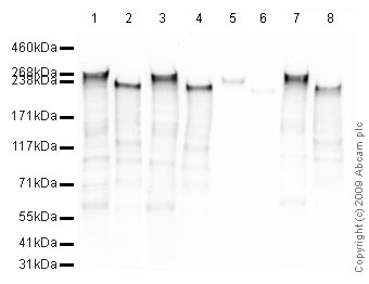
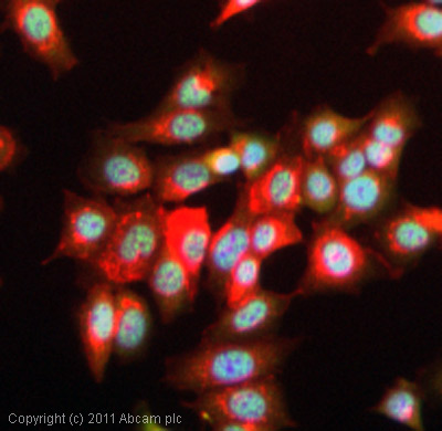
Anti-RNA polymerase II CTD repeat YSPTSPS (phospho S2) antibody - ChIP Grade
| Name | Anti-RNA polymerase II CTD repeat YSPTSPS (phospho S2) antibody - ChIP Grade |
|---|---|
| Supplier | Abcam |
| Catalog | ab5095 |
| Prices | $403.00 |
| Sizes | 100 µg |
| Host | Rabbit |
| Clonality | Polyclonal |
| Isotype | IgG |
| Applications | ELISA IHC-F ChIP ChIP ChIP IHC-P IHC-F ICC/IF ICC/IF DB WB ChIPseq |
| Species Reactivities | Mouse, Rat, Human, Yeast, Xenopus, Arabidopsis thaliana, C. elegans, Drosophila, S. pombe, Broad species |
| Antigen | Synthetic peptide conjugated to KLH derived from within residues 1600 - 1700 of Saccharomyces cerevisiae RNA polymerase II CTD repeat YSPTSPS, phosphorylated at S2 |
| Blocking Peptide | S. cerevisiae RNA polymerase II CTD repeat YSPTSPS (phospho S2) peptide |
| Description | Rabbit Polyclonal |
| Gene | CELE_F36A4.7 |
| Conjugate | Unconjugated |
| Supplier Page | Shop |
Product images
Product References
Systems level-based RNAi screening by high content analysis identifies UBR5 as a - Systems level-based RNAi screening by high content analysis identifies UBR5 as a
Bolt MJ, Stossi F, Callison AM, Mancini MG, Dandekar R, Mancini MA. Oncogene. 2015 Jan 8;34(2):154-64.
Dissecting the function of the adult beta-globin downstream promoter region using - Dissecting the function of the adult beta-globin downstream promoter region using
Barrow JJ, Li Y, Hossain M, Huang S, Bungert J. Nucleic Acids Res. 2014 Apr;42(7):4363-74.
The transcript elongation factor SPT4/SPT5 is involved in auxin-related gene - The transcript elongation factor SPT4/SPT5 is involved in auxin-related gene
Durr J, Lolas IB, Sorensen BB, Schubert V, Houben A, Melzer M, Deutzmann R, Grasser M, Grasser KD. Nucleic Acids Res. 2014 Apr;42(7):4332-47.
A dual role for the histone methyltransferase PR-SET7/SETD8 and histone H4 lysine - A dual role for the histone methyltransferase PR-SET7/SETD8 and histone H4 lysine
Kapoor-Vazirani P, Vertino PM. J Biol Chem. 2014 Mar 14;289(11):7425-37.
Nonproteolytic roles of 19S ATPases in transcription of CIITApIV genes. - Nonproteolytic roles of 19S ATPases in transcription of CIITApIV genes.
Maganti N, Moody TD, Truax AD, Thakkar M, Spring AM, Germann MW, Greer SF. PLoS One. 2014 Mar 13;9(3):e91200.
H3K27me3 and H3K4me3 chromatin environment at super-induced dehydration stress - H3K27me3 and H3K4me3 chromatin environment at super-induced dehydration stress
Liu N, Fromm M, Avramova Z. Mol Plant. 2014 Mar;7(3):502-13.
Diversity of two forms of DNA methylation in the brain. - Diversity of two forms of DNA methylation in the brain.
Chen Y, Damayanti NP, Irudayaraj J, Dunn K, Zhou FC. Front Genet. 2014 Mar 10;5:46.
CpG domains downstream of TSSs promote high levels of gene expression. - CpG domains downstream of TSSs promote high levels of gene expression.
Krinner S, Heitzer AP, Diermeier SD, Obermeier I, Langst G, Wagner R. Nucleic Acids Res. 2014 Apr;42(6):3551-64.
The nucleosomal barrier to promoter escape by RNA polymerase II is overcome by - The nucleosomal barrier to promoter escape by RNA polymerase II is overcome by
Skene PJ, Hernandez AE, Groudine M, Henikoff S. Elife. 2014 Apr 15;3:e02042.
Different gene-specific mechanisms determine the 'revised-response' memory - Different gene-specific mechanisms determine the 'revised-response' memory
Liu N, Ding Y, Fromm M, Avramova Z. Nucleic Acids Res. 2014 May;42(9):5556-66.
