Anti-RNA polymerase II CTD repeat YSPTSPS (phospho S2) antibody - ChIP Grade
| Name | Anti-RNA polymerase II CTD repeat YSPTSPS (phospho S2) antibody - ChIP Grade |
|---|---|
| Supplier | Abcam |
| Catalog | ab5095 |
| Prices | $403.00 |
| Sizes | 100 µg |
| Host | Rabbit |
| Clonality | Polyclonal |
| Isotype | IgG |
| Applications | ELISA IHC-F ChIP ChIP ChIP IHC-P IHC-F ICC/IF ICC/IF DB WB ChIPseq |
| Species Reactivities | Mouse, Rat, Human, Yeast, Xenopus, Arabidopsis thaliana, C. elegans, Drosophila, S. pombe, Broad species |
| Antigen | Synthetic peptide conjugated to KLH derived from within residues 1600 - 1700 of Saccharomyces cerevisiae RNA polymerase II CTD repeat YSPTSPS, phosphorylated at S2 |
| Blocking Peptide | S. cerevisiae RNA polymerase II CTD repeat YSPTSPS (phospho S2) peptide |
| Description | Rabbit Polyclonal |
| Gene | CELE_F36A4.7 |
| Conjugate | Unconjugated |
| Supplier Page | Shop |
Product images
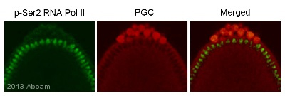 IHC - Wholemount of Caenorhabditis elegans larvae labelling RNA polymerase II CTD repeat YSPTSPS (phospho S2) with ab5095. The sample was incubated with primary antibody (1/500 in PBS + 3% BSA + 0.1% Triton X-100) for 12 hours at 4°C. ab150077, an goat anti-rabbit Alexa Fluor® 488 (1/1000), was used as the secondary antibody.See Abreview
 HeLa or MCF7 cells were fixed with 4% formaldehyde in PEM buffer. The coverslip was incubated in blocking buffer of 5% powdered milk in TBS-T plus 0.02% sodium azide for 1 hour at room temperature. Blocking buffer was removed and primary antibody was added at a dilution of 1/500 and incubated overnight at 4 degrees celsius. The coverslips were then washed 4-5 times with blocking buffer for 5 minutes. Secondary antibody, goat anti-rabbit Alexa 594, was added at a dilution of 1/1000 and incubated at room temperature for one hour. From this point on coverslips were covered with foil to protect them from light. They were washed 5 times with TBS-T and then one time with PEM, for 5 minutes each wash. The coverslips were fixed 10-30 minutes in 4% formaldehyde in PEM buffer, then washed 3 times with PEM buffer for 5 minutes. 0.1M ammonium chloride in PEM buffer was added for 10 minutes to quench auto-florescence, and then slips were washed 2 times for 5 minutes in PEM followed by 3 washes for
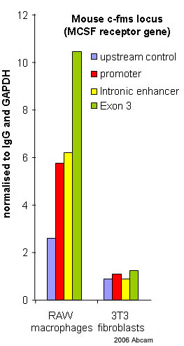 ab5095 at 4ug/ml in ChIP of RAW macrophages. Nuclear cell lysate of mouse RAW macrophages (expressing c-fms) were formaldehyde cross linked and ChIP tested with ab5095. The nuclear preparation was frozen before sonication with a probe sonicator. All buffers used contained protease inhibitors. 3T3 fibroblasts (not expressing c-fms) were used as the negative control.See Abreview
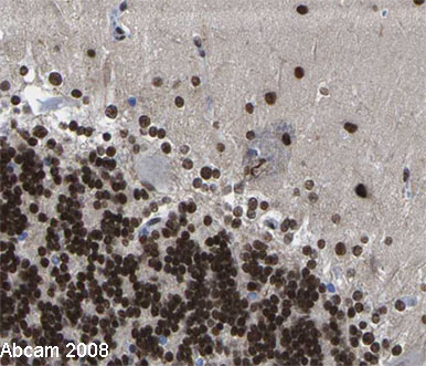 Image courtesy of Human Protein Atlas ab5095 staining in human brain, showing staining of the Purkinje cells (in brown). Paraffin embedded brain tissue was incubated with ab5095 (1:900 dilution) for 30 mins at room temperature. Antigen retrieval was performed by heat induction in citrate buffer pH 6. ab5095 was tested in a tissue microarray (TMA) containing a wide range of normal and cancer tissues as well as a cell microarray consisting of a range of commonly used, well characterised human cell lines. Further results for this antibody can be found at www.proteinatlas.org.
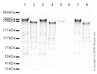 All lanes : Anti-RNA polymerase II CTD repeat YSPTSPS (phospho S2) antibody - ChIP Grade (ab5095) at 1 µg/mlLane 1 : HeLa (Human epithelial carcinoma cell line) Whole Cell Lysate Lane 2 : S.cerevisiae (Y190) Whole Cell Lysate Lane 3 : HeLa (Human epithelial carcinoma cell line) Whole Cell Lysate with S. cerevisiae RNA polymerase II CTD repeat YSPTSPS peptide (ab12795) at 1 µg/mlLane 4 : S.cerevisiae (Y190) Whole Cell Lysate with S. cerevisiae RNA polymerase II CTD repeat YSPTSPS peptide (ab12795) at 1 µg/mlLane 5 : HeLa (Human epithelial carcinoma cell line) Whole Cell Lysate with S. cerevisiae RNA polymerase II CTD repeat YSPTSPS (phospho S2) peptide (ab12793) at 1 µg/mlLane 6 : S.cerevisiae (Y190) Whole Cell Lysate with S. cerevisiae RNA polymerase II CTD repeat YSPTSPS (phospho S2) peptide (ab12793) at 1 µg/mlLane 7 : HeLa (Human epithelial carcinoma cell line) Whole Cell Lysate with Human RNA polymerase II CTD repeat YSPTSPS (phospho S5) peptide (ab18488) at 1 µg/mlLane 8 : S.cerev
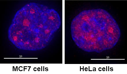
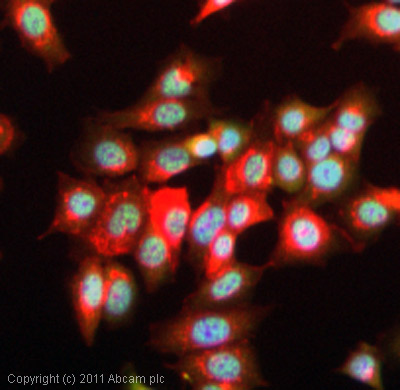 ICC/IF image of ab5095 stained MCF7 cells. The cells were 100% methanol fixed (5 min) and then incubated in 1%BSA / 10% normal goat serum / 0.3M glycine in 0.1% PBS-Tween for 1h to permeabilise the cells and block non-specific protein-protein interactions. The cells were then incubated with the antibody (ab5095, 1µg/ml) overnight at +4°C. The secondary antibody (green) was a goat anti-rabbit DyLight® 488 (IgG - H&L, pre-adsorbed) (ab96899) used at a 1/250 dilution for 1h. Alexa Fluor® 594 WGA was used to label plasma membranes (red) at a 1/200 dilution for 1h. DAPI was used to stain the cell nuclei (blue) at a concentration of 1.43µM.
Product References
Systems level-based RNAi screening by high content analysis identifies UBR5 as a - Systems level-based RNAi screening by high content analysis identifies UBR5 as a
Bolt MJ, Stossi F, Callison AM, Mancini MG, Dandekar R, Mancini MA. Oncogene. 2015 Jan 8;34(2):154-64.
Dissecting the function of the adult beta-globin downstream promoter region using - Dissecting the function of the adult beta-globin downstream promoter region using
Barrow JJ, Li Y, Hossain M, Huang S, Bungert J. Nucleic Acids Res. 2014 Apr;42(7):4363-74.
The transcript elongation factor SPT4/SPT5 is involved in auxin-related gene - The transcript elongation factor SPT4/SPT5 is involved in auxin-related gene
Durr J, Lolas IB, Sorensen BB, Schubert V, Houben A, Melzer M, Deutzmann R, Grasser M, Grasser KD. Nucleic Acids Res. 2014 Apr;42(7):4332-47.
A dual role for the histone methyltransferase PR-SET7/SETD8 and histone H4 lysine - A dual role for the histone methyltransferase PR-SET7/SETD8 and histone H4 lysine
Kapoor-Vazirani P, Vertino PM. J Biol Chem. 2014 Mar 14;289(11):7425-37.
Nonproteolytic roles of 19S ATPases in transcription of CIITApIV genes. - Nonproteolytic roles of 19S ATPases in transcription of CIITApIV genes.
Maganti N, Moody TD, Truax AD, Thakkar M, Spring AM, Germann MW, Greer SF. PLoS One. 2014 Mar 13;9(3):e91200.
H3K27me3 and H3K4me3 chromatin environment at super-induced dehydration stress - H3K27me3 and H3K4me3 chromatin environment at super-induced dehydration stress
Liu N, Fromm M, Avramova Z. Mol Plant. 2014 Mar;7(3):502-13.
Diversity of two forms of DNA methylation in the brain. - Diversity of two forms of DNA methylation in the brain.
Chen Y, Damayanti NP, Irudayaraj J, Dunn K, Zhou FC. Front Genet. 2014 Mar 10;5:46.
CpG domains downstream of TSSs promote high levels of gene expression. - CpG domains downstream of TSSs promote high levels of gene expression.
Krinner S, Heitzer AP, Diermeier SD, Obermeier I, Langst G, Wagner R. Nucleic Acids Res. 2014 Apr;42(6):3551-64.
The nucleosomal barrier to promoter escape by RNA polymerase II is overcome by - The nucleosomal barrier to promoter escape by RNA polymerase II is overcome by
Skene PJ, Hernandez AE, Groudine M, Henikoff S. Elife. 2014 Apr 15;3:e02042.
Different gene-specific mechanisms determine the 'revised-response' memory - Different gene-specific mechanisms determine the 'revised-response' memory
Liu N, Ding Y, Fromm M, Avramova Z. Nucleic Acids Res. 2014 May;42(9):5556-66.