Anti-ROC1 antibody
| Name | Anti-ROC1 antibody |
|---|---|
| Supplier | Abcam |
| Catalog | ab2977 |
| Prices | $390.00 |
| Sizes | 500 µl |
| Host | Rabbit |
| Clonality | Polyclonal |
| Isotype | IgG |
| Applications | ICC/IF ICC/IF IHC-P IP WB |
| Species Reactivities | Human, Mouse |
| Antigen | Synthetic peptide: CPLDNREWEFQKYGH , corresponding to amino acids 97-108 of Human ROC1 |
| Description | Rabbit Polyclonal |
| Gene | RBX1 |
| Conjugate | Unconjugated |
| Supplier Page | Shop |
Product images
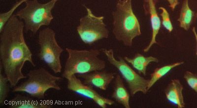 ICC/IF image of ab2977 stained HeLa cells. The cells were 4% PFA fixed (10 min) and then incubated in 1%BSA / 10% normal goat serum / 0.3M glycine in 0.1% PBS-Tween for 1h to permeabilise the cells and block non-specific protein-protein interactions. The cells were then incubated with the antibody (ab2977, 5µg/ml) overnight at +4°C. The secondary antibody (green)ÿwas Alexa Fluor© 488 goat anti-rabbit IgG (H+L) used at a 1/1000 dilution for 1h. Alexa Fluor© 594 WGA was used to label plasma membranes (red) at a 1/200 dilution for 1h. DAPI was used to stain the cell nuclei (blue) at a concentration of 1.43µM.
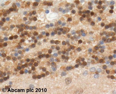 ab2977 (2µg/ml) staining ROC1 in human Brain: Cerebellum using an automated system (DAKO Autostainer Plus). Using this protocol there is cytoplasmic and nuclear staining.Sections were rehydrated and antigen retrieved with the Dako 3-in-1 AR buffer citrate pH 6.0 in a DAKO PT Link. Slides were peroxidase blocked in 3% H2O2 in methanol for 10 minutes. They were then blocked with Dako Protein block for 10 minutes (containing casein 0.25% in PBS) then incubated with primary antibody for 20 minutes and detected with Dako Envision Flex amplification kit for 30 minutes. Colorimetric detection was completed with Diaminobenzidine for 5 minutes. Slides were counterstained with Haematoxylin and coverslipped under DePeX. Please note that, for manual staining, optimization of primary antibody concentration and incubation time is recommended. Signal amplification may be required.
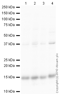 All lanes : Anti-ROC1 antibody (ab2977) at 1 µg/mlLane 1 : HeLa (Human epithelial carcinoma cell line) Whole Cell LysateLane 2 : Jurkat (Human T cell lymphoblast-like cell line) Whole Cell Lysate Lane 3 : HepG2 (Human hepatocellular liver carcinoma cell line) Whole Cell LysateLane 4 : MCF7 (Human breast adenocarcinoma cell line) Whole Cell LysateLysates/proteins at 10 µg per lane.SecondaryGoat Anti-Rabbit IgG H&L (HRP) preadsorbed (ab97080) at 1/5000 dilutiondeveloped using the ECL techniquePerformed under reducing conditions.
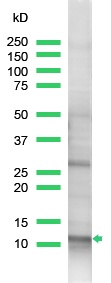 Anti-ROC1 antibody (ab2977) at 1/25 dilution + HeLa cell lysate
Product References
Endoplasmic reticulum stress induces a caspase-dependent N-terminal cleavage of - Endoplasmic reticulum stress induces a caspase-dependent N-terminal cleavage of
Shteingart S, Hadar R, Cohen I, Ravid T, Tirosh B. J Biol Chem. 2012 Sep 7;287(37):31223-32.
SCCRO (DCUN1D1) promotes nuclear translocation and assembly of the neddylation E3 - SCCRO (DCUN1D1) promotes nuclear translocation and assembly of the neddylation E3
Huang G, Kaufman AJ, Ramanathan Y, Singh B. J Biol Chem. 2011 Mar 25;286(12):10297-304.
RNAi screen of Salmonella invasion shows role of COPI in membrane targeting of - RNAi screen of Salmonella invasion shows role of COPI in membrane targeting of
Misselwitz B, Dilling S, Vonaesch P, Sacher R, Snijder B, Schlumberger M, Rout S, Stark M, von Mering C, Pelkmans L, Hardt WD. Mol Syst Biol. 2011 Mar 15;7:474.
VHL type 2B mutations retain VBC complex form and function. - VHL type 2B mutations retain VBC complex form and function.
Hacker KE, Lee CM, Rathmell WK. PLoS One. 2008;3(11):e3801.