Anti-Rubicon antibody - N-terminal (ab173930) at 1/50 dilution + MCF7 cell lysate at 35 µg

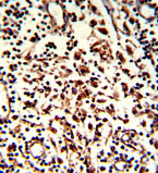
Immunohistochemical analysis of formalin-fixed, paraffin-embedded Human lymph tissue labeling Rubicon with ab173930 at 1/50 dilution, which was peroxidase-conjugated to the secondary antibody, followed by DAB staining.

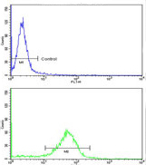
Flow cytometric analysis of MDA-231 cells labeling Rubicon with ab173930 at 1/10 dilution (bottom histogram), compared to negative control cells (top histogram). FITC-conjugated goat-anti-rabbit secondary antibodies were used for the analysis.

Immunofluorescent analysis of U251 cells labeling Rubicon with ab173930 at 1/100 dilution. U251 cells were treated with Chloroquine (50 μM,16h), then fixed with 4% PFA (20 min), permeabilized with Triton X-100 (0.2%, 30 min). Cells were then incubated with ab173930 (1/100, 2 h at room temperature). For secondary antibody, Alexa Fluor® 488 conjugated donkey anti-rabbit antibody (green) was used (1/1000, 1h). Nuclei were counterstained with Hoechst 33342 (blue) (10 μg/ml, 5 min).


