![All lanes : Anti-SET antibody [EPR12973] (ab181990) at 1/100000 dilutionLane 1 : Jurkat cell lysateLane 2 : 293T cell lysateLane 3 : Hela cell lysateLane 4 : HepG2 cell lysateLysates/proteins at 20 µg per lane.SecondaryGoat Anti-Rabbit IgG, (H+L), Peroxidase conjugated at 1/1000 dilution](http://www.bioprodhub.com/system/product_images/ab_products/2/sub_4/28863_ab181990-212554-ab1819901.jpg)
All lanes : Anti-SET antibody [EPR12973] (ab181990) at 1/100000 dilutionLane 1 : Jurkat cell lysateLane 2 : 293T cell lysateLane 3 : Hela cell lysateLane 4 : HepG2 cell lysateLysates/proteins at 20 µg per lane.SecondaryGoat Anti-Rabbit IgG, (H+L), Peroxidase conjugated at 1/1000 dilution
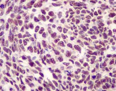
Immunohistochemical analysis of paraffin-embedded Human nephroblastoma tissue labeling SET with ab181990 at a 1/250 dilution. Prediluted (ready to use) HRP Polymer for Rabbit IgG was used as a secondary antibody. Counter stain: Hematoxylin
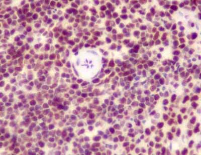
Immunohistochemical analysis of paraffin-embedded rat spleen tissue labeling SET with ab181990 at a 1/250 dilution. Prediluted (ready to use) HRP Polymer for Rabbit IgG was used as a secondary antibody. Counter stain: Hematoxylin
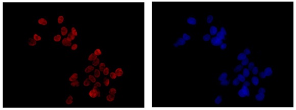
Immunofluorescent analysis of 4% paraformaldehyde fixed HepG2 cell line staining SET using ab181990 at 1/1000 dilution. A Dylight 555 conjugated Goat anti rabbit IgG at 1/200 dilution was used for secondary antibody detection. Counter stain: Dapi.
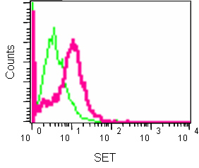
Flow cytometric analysis of 2% paraformaldehyde-fixed Hela cells labeling SET using ab181990 at a 1/10 dilution, or a rabbit monoclonal IgG isotype control. Secondary antibody used was goat anti rabbit IgG (FITC) at a 1/150 dilution.
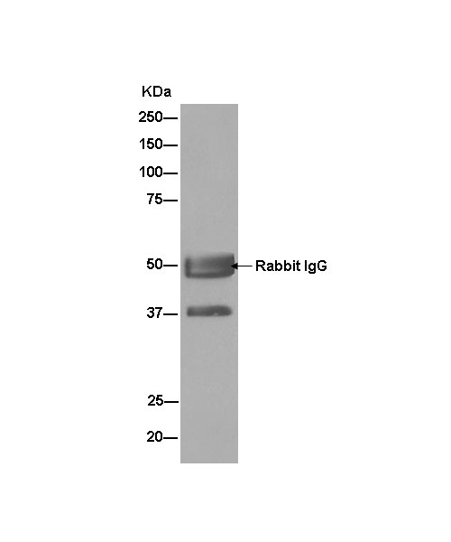
Immunoprecipitation of Jurkat cell lysate labeling SET using ab181990 at a 1/30 dilution (lane 1). Lane 2 shows the negative control. A goat anti-rabbit IgG, (H+L), peroxidase conjugated secondary antibody was used at a dilution of 1/1000. Blocking/dilution buffer and concentration: 5% NFDM/TBST.