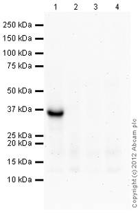
All lanes : Anti-SLUG antibody (ab106077) at 1 µg/mlLane 1 : MCF7 cell lysate overexpressing SLUG proteinLane 2 : MCF7 cell lysate overexpressing SNAIL proteinLane 3 : MCF7 cell lysate transfected with vector control plasmidLane 4 : Untransfected MCF7 cell lysateLysates/proteins at 10 µg per lane.SecondaryGoat Anti-Rabbit IgG H&L (HRP) preadsorbed (ab97080) at 1/5000 dilutiondeveloped using the ECL techniquePerformed under reducing conditions.

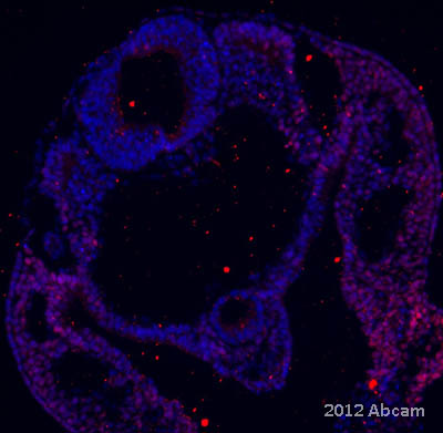
ab106077 staining SLUG on an E9.5 mouse embryo section by immunohistochemistry. The tissue was paraformaldehyde fixed and permeabilized in PBST prior to blocking in NOVOCASTRA protein block for 30 minutes at 25°C. The primary antibody was diluted 1/25 and incubated with the sample for 48 hour at 4°C. An Alexa Fluor 555 conjugated donkey anti-rabbit antibody, diluted 1/1000, was used as the secondary. Nuclear staining shown with DAPI.See Abreview

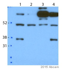
All lanes : Anti-SLUG antibody (ab106077) at 1/2000 dilutionLane 1 : MDA-MB-231 whole cell lysateLane 2 : MDA-MB-231 whole cell lysate - sh Slug 1Lane 3 : MDA-MB-231 whole cell lysate - sh Slug 2Lane 4 : MDA-MB-231 whole cell lysate - sh Snail 1Lysates/proteins at 30 µg per lane.SecondaryHRP-conjugated goat anti-rabbit IgG polyclonal at 1/5000 dilutiondeveloped using the ECL techniquePerformed under reducing conditions.


