Anti-SQSTM1 / p62 antibody
| Name | Anti-SQSTM1 / p62 antibody |
|---|---|
| Supplier | Abcam |
| Catalog | ab56416 |
| Prices | $401.00 |
| Sizes | 100 µg |
| Host | Mouse |
| Clonality | Monoclonal |
| Isotype | IgG2a |
| Applications | IHC-P WB ICC/IF ICC/IF FC IHC-F |
| Species Reactivities | Mouse, Rat, Human, Monkey, Hamster |
| Antigen | Recombinant full length protein, corresponding to amino acids 1-441 of Human SQSTM1/ p62 |
| Description | Mouse Monoclonal |
| Gene | SQSTM1 |
| Conjugate | Unconjugated |
| Supplier Page | Shop |
Product images
Product References
Ambroxol improves lysosomal biochemistry in glucocerebrosidase mutation-linked - Ambroxol improves lysosomal biochemistry in glucocerebrosidase mutation-linked
McNeill A, Magalhaes J, Shen C, Chau KY, Hughes D, Mehta A, Foltynie T, Cooper JM, Abramov AY, Gegg M, Schapira AH. Brain. 2014 May;137(Pt 5):1481-95.
Cellular consequences of oxidative stress in riboflavin responsive multiple - Cellular consequences of oxidative stress in riboflavin responsive multiple
Cornelius N, Corydon TJ, Gregersen N, Olsen RK. Hum Mol Genet. 2014 Aug 15;23(16):4285-301.
Important role of autophagy in endothelial cell response to ionizing radiation. - Important role of autophagy in endothelial cell response to ionizing radiation.
Kalamida D, Karagounis IV, Giatromanolaki A, Koukourakis MI. PLoS One. 2014 Jul 10;9(7):e102408.
Fasting increases human skeletal muscle net phenylalanine release and this is - Fasting increases human skeletal muscle net phenylalanine release and this is
Vendelbo MH, Moller AB, Christensen B, Nellemann B, Clasen BF, Nair KS, Jorgensen JO, Jessen N, Moller N. PLoS One. 2014 Jul 14;9(7):e102031.
Prognostic significance of p62/SQSTM1 subcellular localization and LC3B in oral - Prognostic significance of p62/SQSTM1 subcellular localization and LC3B in oral
Liu JL, Chen FF, Lung J, Lo CH, Lee FH, Lu YC, Hung CH. Br J Cancer. 2014 Aug 26;111(5):944-54.
Targeting aPKC disables oncogenic signaling by both the EGFR and the - Targeting aPKC disables oncogenic signaling by both the EGFR and the
Kusne Y, Carrera-Silva EA, Perry AS, Rushing EJ, Mandell EK, Dietrich JD, Errasti AE, Gibbs D, Berens ME, Loftus JC, Hulme C, Yang W, Lu Z, Aldape K, Sanai N, Rothlin CV, Ghosh S. Sci Signal. 2014 Aug 12;7(338):ra75.
Autophagy promotes paclitaxel resistance of cervical cancer cells: involvement of - Autophagy promotes paclitaxel resistance of cervical cancer cells: involvement of
Peng X, Gong F, Chen Y, Jiang Y, Liu J, Yu M, Zhang S, Wang M, Xiao G, Liao H. Cell Death Dis. 2014 Aug 14;5:e1367.
Interaction of SQSTM1 with the motor protein dynein--SQSTM1 is required for - Interaction of SQSTM1 with the motor protein dynein--SQSTM1 is required for
Calderilla-Barbosa L, Seibenhener ML, Du Y, Diaz-Meco MT, Moscat J, Yan J, Wooten MW, Wooten MC. J Cell Sci. 2014 Sep 15;127(Pt 18):4052-63.
Mice deficient in Epg5 exhibit selective neuronal vulnerability to degeneration. - Mice deficient in Epg5 exhibit selective neuronal vulnerability to degeneration.
Zhao H, Zhao YG, Wang X, Xu L, Miao L, Feng D, Chen Q, Kovacs AL, Fan D, Zhang H. J Cell Biol. 2013 Mar 18;200(6):731-41.
.

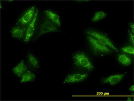
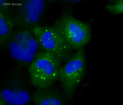
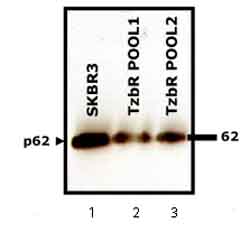
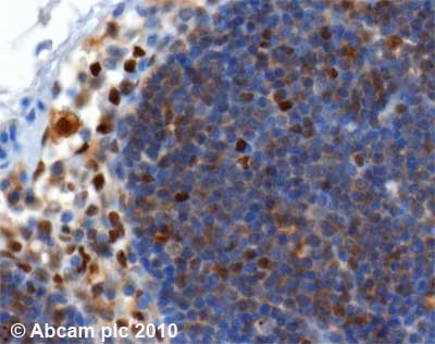
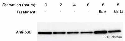
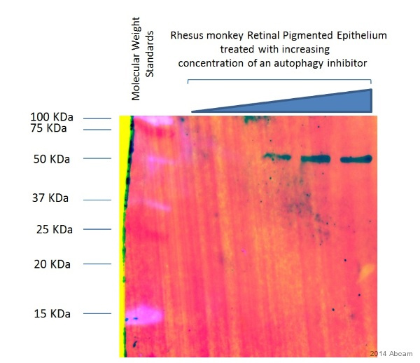
![Overlay histogram showing HeLa cells stained with ab56416 (red line). The cells were fixed with 80% methanol (5 min) and then permeabilized with 0.1% PBS-Tween for 20 min. The cells were then incubated in 1x PBS / 10% normal goat serum / 0.3M glycine to block non-specific protein-protein interactions followed by the antibody (ab56416, 0.5µg/1x106 cells) for 30 min at 22ºC. The secondary antibody used was DyLight® 488 goat anti-mouse IgG (H+L) (ab96879) at 1/500 dilution for 30 min at 22ºC. Isotype control antibody (black line) was mouse IgG2a [ICIGG2A] (ab91361, 1µg/1x106 cells) used under the same conditions. Acquisition of >5,000 events was performed.](http://www.bioprodhub.com/system/product_images/ab_products/2/sub_5/4706_SQSTM1-p62-Primary-antibodies-ab56416-53.jpg)
