SQSTM1

Gene Symbol SQSTM1
Entrez Gene 8878
Alt Symbol A170, OSIL, PDB3, ZIP3, p60, p62, p62B
Species Human
Gene Type protein-coding
Description sequestosome 1
Other Description EBI3-associated protein of 60 kDa|EBI3-associated protein p60|EBIAP|oxidative stress induced like|phosphotyrosine independent ligand for the Lck SH2 domain p62|phosphotyrosine-independent ligand for the Lck SH2 domain of 62 kDa|sequestosome-1|ubiquitin-binding protein p62
Swissprots Q13501 Q9BUV7 B3KUW5 A6NFN7 Q13446 Q9UEU1 Q9BVS6 B2R661
Accessions AAC64516 EAW53786 EAW53787 Q13501 AI333228 AK025146 AK093109 AK094484 AK096241 AK096257 AK098077 BAG53577 AK226167 AK293509 BAG56993 AK304877 BAG65614 AK312451 BAG35358 BC000951 AAH00951 BC001874 AAH01874 BC003139 AAH03139 BC005857 BC017222 AAH17222 BC019111 AAH19111 BC050631 BI549805 BM800056 BU625974 DA541885 DC398116 DQ581944 DQ892878 ABM83804 EU176717 ABW03518 U41806 AAA93299 U46751 AAC52070 XM_011534683 XP_011532985 XM_011534684 XP_011532986 NM_001142298 NP_001135770 NM_001142299 NP_001135771 NM_003900 NP_003891
Function Autophagy receptor that interacts directly with both the cargo to become degraded and an autophagy modifier of the MAP1 LC3 family. Required both for the formation and autophagic degradation of polyubiquitin-containing bodies, called ALIS (aggresome-like induced structures) and links ALIS to the autophagic machinery. Involved in midbody ring degradation. May regulate the activation of NFKB1 by TNF-alpha, nerve growth factor (NGF) and interleukin- 1. May play a role in titin/TTN downstream signaling in muscle cells. May regulate signaling cascades through ubiquitination. Adapter that mediates the interaction between TRAF6 and CYLD (By similarity). May be involved in cell differentiation, apoptosis, immune response and regulation of K(+) channels. {ECO:0000250, ECO:0000269|PubMed:10356400, ECO:0000269|PubMed:10747026, ECO:0000269|PubMed:11244088, ECO:0000269|PubMed:12471037, ECO:0000269|PubMed:15340068, ECO:0000269|PubMed:15802564, ECO:0000269|PubMed:15911346, ECO:0000269|PubMed:15953362
Subcellular Location Cytoplasm. Late endosome. Lysosome. Cytoplasmic vesicle, autophagosome. Nucleus. Endoplasmic reticulum. Cytoplasm, P-body. Note=Sarcomere (By similarity). In cardiac muscles localizes to the sarcomeric band (By similarity). Commonly found in inclusion bodies containing polyubiquitinated protein aggregates. In neurodegenerative diseases, detected in Lewy bodies in Parkinson disease, neurofibrillary tangles in Alzheimer disease, and HTT aggregates in Huntington disease. In protein aggregate diseases of the liver, found in large amounts in Mallory bodies of alcoholic and nonalcoholic steatohepatitis, hyaline bodies in hepatocellular carcinoma, and in SERPINA1 aggregates. Enriched in Rosenthal fibers of pilocytic astrocytoma. In the cytoplasm, observed in both membrane-free ubiquitin- containing protein aggregates (sequestosomes) and membrane- surrounded autophagosomes. Colocalizes with TRIM13 in the perinuclear endoplasmic reticulum. Co-localizes with TRIM5 in the cytoplasmic bodies. {ECO
Tissue Specificity Ubiquitously expressed. {ECO:0000269|PubMed:8650207}.
Top Pathways Osteoclast differentiation

SQSTM1/p62 (D1D9E3) Rabbit mAb (Alexa Fluor ® 488 Conjugate) - 8833 from Cell Signaling Technology

Prices $299.00
Sizes 100 µl (50 tests)
Host Rabbit
Clonality Monoclonal
Clone D1D9E3
Confocal immunofluorescent analysis of HeLa cells, untreated (left) or treated with bafilomycin A (100 nM, 18 hr; right), using SQSTM1/p62 (D1D9E3) Rabbit mAb (Alexa Fluor ® 488 Conjugate) (green). Actin filaments were labeled with DY-554 phalloidin. Blue pseudocolor = DRAQ5 ® #4084 (fluorescent DNA dye).

Phospho-SQSTM1/p62 (Thr269/Ser272) Antibody - 13121 from Cell Signaling Technology

Prices $287.00
Sizes 100 µl (10 western blots)
Host Rabbit
Clonality Polyclonal
Western blot analysis of extracts from A549 cells, untreated (-) or treated with Roscovitine #9885 (20 μM, overnight; +), using Phospho-SQSTM1/p62 (Thr269/Ser272) Antibody (upper), SQSTM1/p62 (D5E2) Rabbit mAb #8025 (middle) or β-Actin (D6A8) Rabbit mAb #8457 (lower).

Phospho-SQSTM1/p62 (Ser403) Antibody - 14354 from Cell Signaling Technology

Prices $279.00
Sizes 100 µl (10 western blots)
Host Rabbit
Clonality Polyclonal
Western blot analysis of extracts from THP-1 cells differentiated with TPA #4174 (20 ng/ml, overnight; -) followed by transfection with poly(deoxyadenylic-deoxythymidylic) acid (poly(dA:dT); 2 μg/ml, 7 hr; +) using Phospho-SQSTM1/p62 (Ser403) Antibody (upper) or total SQSTM1/p62 (D5E2) Rabbit mAb #8025 (lower).

SQSTM1/p62 (D10E10) Rabbit mAb (IF Preferred) - 7695 from Cell Signaling Technology

Prices $246.00
Sizes 100 µl (400 tests)
Host Rabbit
Clonality Monoclonal
Clone D10E10
Confocal immunofluorescent analysis of HeLa cells, untreated (left) or treated with bafilomycin A (100 nM, 18 hr; right), using SQSTM1/p62 (D10E10) Rabbit mAb (IF Preferred) (green). Actin filaments were labeled with DY-554 phalloidin. Blue pseudocolor = DRAQ5 ® #4084 (fluorescent DNA dye).

SQSTM1/p62 Antibody - 5114 from Cell Signaling Technology

Prices $99.00, $246.00
Sizes 20 µl (2 western blots), 100 µl (10 western blots)
Host Rabbit
Clonality Polyclonal
Western blot analysis of extracts from various cell lines using SQSTM1/p62 Antibody.

SQSTM1/p62 (D5E2) Rabbit mAb - 8025 from Cell Signaling Technology

Prices $99.00, $246.00
Sizes 20 µl (2 western blots), 100 µl (10 western blots)
Host Rabbit
Clonality Monoclonal
Clone D5E2
Western blot analysis of extracts from HeLa cells, transfected with 100 nM SignalSilence ® Control siRNA (Unconjugated) #6568 (-), SignalSilence ® SQSTM1/p62 siRNA I #6394 (+) or SignalSilence ® SQSTM1/p62 siRNA II #6399 (+), using SQSTM1/p62 (D5E2) Rabbit mAb (upper) or α-Tubulin (11H10) Rabbit mAb #2125 (lower). The SQSTM1/p62 (D5E2) Rabbit mAb confirms silencing of SQSTM1/p62 expression, while the α-Tubulin (11H10) Rabbit mAb is used as a loading control.

SQSTM1 (P-15) - sc-10117 from Santa Cruz Biotechnology

Prices $279.00
Sizes 200 µg/ml
Host Goat
Clonality Polyclonal
SQSTM1 (P-15): sc-10117. Western blot analysis of SQSTM1 expression in HeLa whole cell lysate.

SQSTM1 (H-290) - sc-25575 from Santa Cruz Biotechnology

Prices $279.00
Sizes 200 µg/ml
Host Rabbit
Clonality Polyclonal
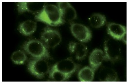
SQSTM1 (H-290): sc-25575. Immunofluorescence staining of methanol-fixed HeLa cells showing cytoplasmic localization.

SQSTM1 (D-3) - sc-28359 from Santa Cruz Biotechnology

Prices $279.00
Sizes 200 µg/ml
Host Mouse
Clonality Monoclonal
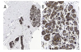
SQSTM1 (D-3): sc-28359. Immunoperoxidase staining of formalin fixed, paraffin-embedded human breast tissue showing cytoplasmic staining of glandular cells at low (A) and high (B) magnification. Kindly provided by The Swedish Human Protein Atlas (HPA) program.

SQSTM1 (A-6) - sc-48402 from Santa Cruz Biotechnology

Prices $279.00
Sizes 200 µg/ml
Host Mouse
Clonality Monoclonal
SQSTM1 (A-6): sc-48402. Immunofluorescence staining of methanol-fixed HeLa cells showing cytoplasmic localization.

Anti-SQSTM1 / p62 antibody - ab56416 from Abcam

Prices $401.00
Sizes 100 µg
Host Mouse
Clonality Monoclonal
Western blot against tagged recombinant protein immunogen using ab56416 SQSTM1 / p62 antibody at 1ug/ml.

Anti-SQSTM1 / p62 antibody - ab91526 from Abcam

Prices $401.00
Sizes 100 µg
Host Rabbit
Clonality Polyclonal
Lane 1 :  Anti-SQSTM1 / p62 antibody (ab91526) at 1 µg/mlLane 2 :  Anti-SQSTM1 / p62 antibody (ab91526) at 2 µg/mlLane 1 : Human spleen tissue lysateLane 2 : Human spleen tissue lysateLysates/proteins at 15 µg per lane.

Anti-SQSTM1 / p62 antibody - ab64134 from Abcam

Host Rabbit
Clonality Polyclonal
IHC image of ab64134 staining in human pancreas formalin fixed paraffin embedded tissue section, performed on a Leica BondTM system using the standard protocol F. The section was pre-treated using heat mediated antigen retrieval with sodium citrate buffer (pH6, epitope retrieval solution 1) for 20 mins. The section was then incubated with ab64134, 5µg/ml, for 15 mins at room temperature and detected using an HRP conjugated compact polymer system. DAB was used as the chromogen. The section was then counterstained with haematoxylin and mounted with DPX.For other IHC staining systems (automated and non-automated) customers should optimize variable parameters such as antigen retrieval conditions, primary antibody concentration and antibody incubation times.

Anti-SQSTM1 / p62 antibody - ab31545 from Abcam

Prices $382.00
Sizes 100 µl
Host Sheep
Clonality Polyclonal
All lanes : Anti-SQSTM1 / p62 antibody (ab31545) at 1/500 dilutionLane 1 : A549 (Human lung adenocarcinoma epithelial cell line) Whole Cell LysateLane 2 : MCF7 (Human breast adenocarcinoma cell line) Whole Cell LysateLane 3 : HepG2 (Human hepatocellular liver carcinoma cell line) Whole Cell Lysate Lysates/proteins at 10 µg per lane.SecondaryRabbit polyclonal Secondary Antibody to Sheep IgG - H&L (HRP) at 1/3000 dilutiondeveloped using the ECL techniquePerformed under reducing conditions.

Anti-SQSTM1 / p62 antibody - ab155686 from Abcam

Prices $376.00
Sizes 100 µl
Host Rabbit
Clonality Polyclonal
Anti-SQSTM1 / p62 antibody (ab155686) at 1/1000 dilution + A549 whole cell lysate at 30 µg

Anti-SQSTM1 / p62 antibody - ab76339 from Abcam

Host Sheep
Clonality Polyclonal

Anti-SQSTM1 / p62 antibody [ EPR4844] (DyLight® 488) - ab139895 from Abcam

Prices $371.00
Sizes 100 µl
Host Rabbit
Clonality Monoclonal
Clone EPR4844
Immunofluorescent analysis of HeLa cells labelling SQSTM1 / p62 with ab139895 at 1/100 dilution (green); DAPI staining (blue).

Anti-SQSTM1 / p62 antibody [1B2] - ab118275 from Abcam

Prices $376.00
Sizes 100 µl
Host Mouse
Clonality Monoclonal
Clone 1B2
All lanes : Anti-SQSTM1 / p62 antibody [1B2] (ab118275) at 1/500 dilutionLane 1 : Non transfected cell lysateLane 2 : SQSTM1 / p62 transfected HEK293T cells Lysates/proteins at 5 µg per lane.

Anti-SQSTM1 / p62 antibody [5D5] - ab119409 from Abcam

Prices $376.00
Sizes 100 µl
Host Mouse
Clonality Monoclonal
Clone 5D5
All lanes : Anti-SQSTM1 / p62 antibody [5D5] (ab119409) (1/500)Lane 1 : HEK293T cells transfected with control cDNA for 48 hoursLane 2 : HEK293T cells transfected with SQSTM1 / p62 cDNA for 48 hoursLysates/proteins at 5 µg per lane.

Anti-SQSTM1 / p62 antibody (Biotin) - ab191819 from Abcam

Prices $359.00
Sizes 100 µl
Host Rabbit
Clonality Polyclonal

Anti-SQSTM1 / p62 antibody [EPR4844] (Alexa Fluor® 647) - ab194721 from Abcam

Prices $425.00
Sizes 100 µl
Host Rabbit
Clonality Monoclonal
Clone EPR4844
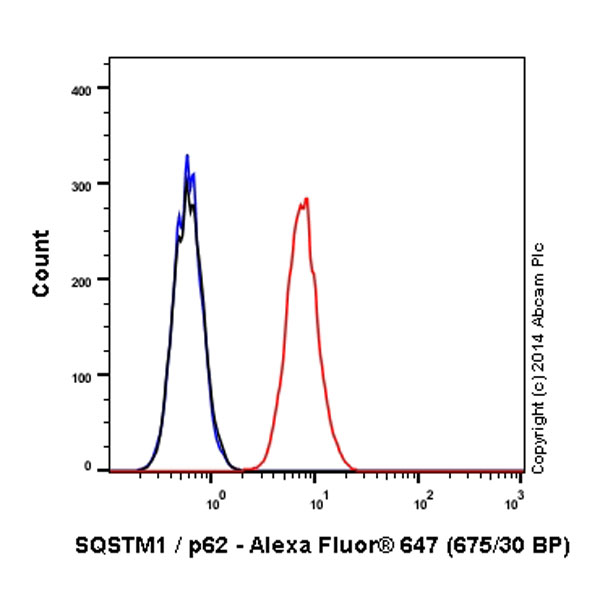
Overlay histogram showing MCF7 cells stained with ab194721 (red line). The cells were fixed with 4% formaldehyde (10 min) and then permeabilized with 0.1% PBS-Tween for 20 min. The cells were then incubated in 1x PBS / 10% normal goat serum / 0.3M glycine to block non-specific protein-protein interactions followed by the antibody (ab194721, 1/500 dilution) for 30 min at 22°C. Isotype control antibody (black line) was rabbit IgG (monoclonal) Alexa Fluor® 647 used at the same concentration and conditions as the primary antibody. Unlabelled sample (blue line) was also used as a control.Acquisition of >5,000 events were collected using a solid-state 25mW red diode laser (635 nm) and 675/30 bandpass filter.This antibody gave a positive signal in MCF7 fixed with 80% methanol (5 min)/permeabilized with 0.1% PBS-Tween for 20 min used under the same conditions.

Anti-SQSTM1 / p62 antibody [EPR4844] (HRP) - ab194720 from Abcam

Prices $425.00
Sizes 100 µl
Host Rabbit
Clonality Monoclonal
Clone EPR4844
All lanes : Anti-SQSTM1 / p62 antibody [EPR4844] (HRP) (ab194720) at 1/1000 dilutionLane 1 : MCF7 (Human breast adenocarcinoma cell line) Whole Cell LysateLane 2 : HeLa (Human epithelial carcinoma cell line) Whole Cell Lysate (ab150035)Lysates/proteins at 10 µg per lane.developed using the ECL techniquePerformed under reducing conditions.

Anti-SQSTM1 / p62 antibody [EPR4844] - Autophagosome Marker - ab109012 from Abcam

Prices $385.00
Sizes 100 µl
Host Rabbit
Clonality Monoclonal
Clone EPR4844
Overlay histogram showing HeLa cells stained with ab109012 (red line). The cells were fixed with 80% methanol (5 min) and then permeabilized with 0.1% PBS-Tween for 20 min. The cells were then incubated in 1x PBS / 10% normal goat serum / 0.3M glycine to block non-specific protein-protein interactions followed by the antibody (ab109012, 1/100 dilution) for 30 min at 22°C. The secondary antibody used was Alexa Fluor® 488 goat anti-rabbit IgG (H+L) (ab150077) at 1/2000 dilution for 30 min at 22°C. Isotype control antibody (black line) was rabbit IgG (monoclonal) (1μg/1x106 cells) used under the same conditions. Unlabelled sample (blue line) was also used as a control. Acquisition of >5,000 events were collected using a 20mW Argon ion laser (488nm) and 525/30 bandpass filter.

Anti-SQSTM1 / p62 antibody [EPR4844] - Autophagosome Marker (Alexa Fluor® 488) - ab185015 from Abcam

Prices $431.00
Sizes 100 µl
Host Rabbit
Clonality Monoclonal
Clone EPR4844
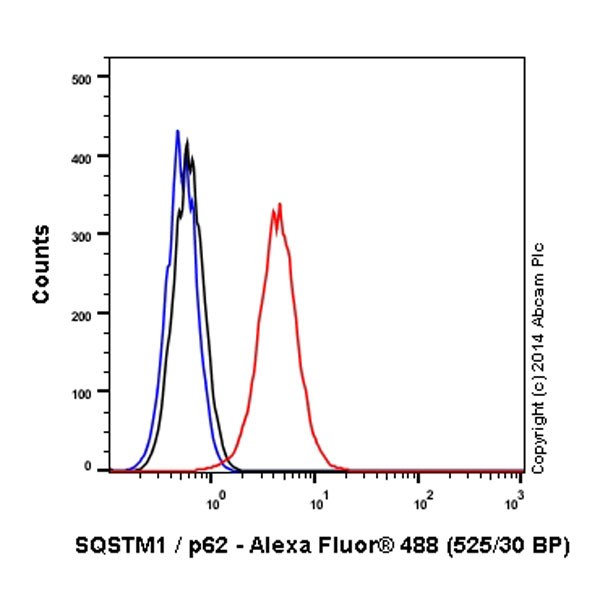
Overlay histogram showing HeLa cells stained with ab185015 (red line). The cells were fixed with 4% formaldehyde (10 min) and then permeabilized with 0.1% PBS-Tween for 20 min. The cells were then incubated in 1x PBS / 10% normal goat serum / 0.3M glycine to block non-specific protein-protein interactions followed by the antibody (ab185015, 1/500 dilution) for 30 min at 22°C. Isotype control antibody (black line) was rabbit IgG (monoclonal) Alexa Fluor® 488 used at the same concentration and conditions as the primary antibody. Unlabelled sample (blue line) was also used as a control.Acquisition of >5,000 events were collected using a 20mW Argon ion laser (488nm) and 525/30 bandpass filter.This antibody gave a positive signal in HeLa fixed with 80% methanol (5 min)/permeabilized with 0.1% PBS-Tween for 20 min used under the same conditions.

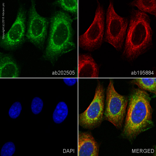
Anti-SQSTM1 / p62 antibody [EPR4844] - Autophagosome Marker (Alexa Fluor® 568) - ab202505 from Abcam

Prices $425.00
Sizes 100 µl
Host Rabbit
Clonality Monoclonal
Clone EPR4844
ab202505 staining SQSTM1/p62 in MCF7 cells. The cells were fixed with 100% methanol (5min), permeabilized with 0.1% Triton X-100 for 5 minutes and then blocked with 1% BSA/10% normal goat serum/0.3M glycine in 0.1% PBS-Tween for 1h. The cells were then incubated overnight at +4°C with ab202505 at 1/200 dilution (shown in green) and ab195884, Rat monoclonal to Tubulin (Alexa Fluor® 647), at 1/250 dilution (shown in red). Nuclear DNA was labelled with DAPI (shown in blue).Image was taken with a confocal microscope (Leica-Microsystems, TCS SP8).This product also gave a positive signal under the same testing conditions in MCF7 cells fixed with 4% formaldehyde (10 min).
Search more